
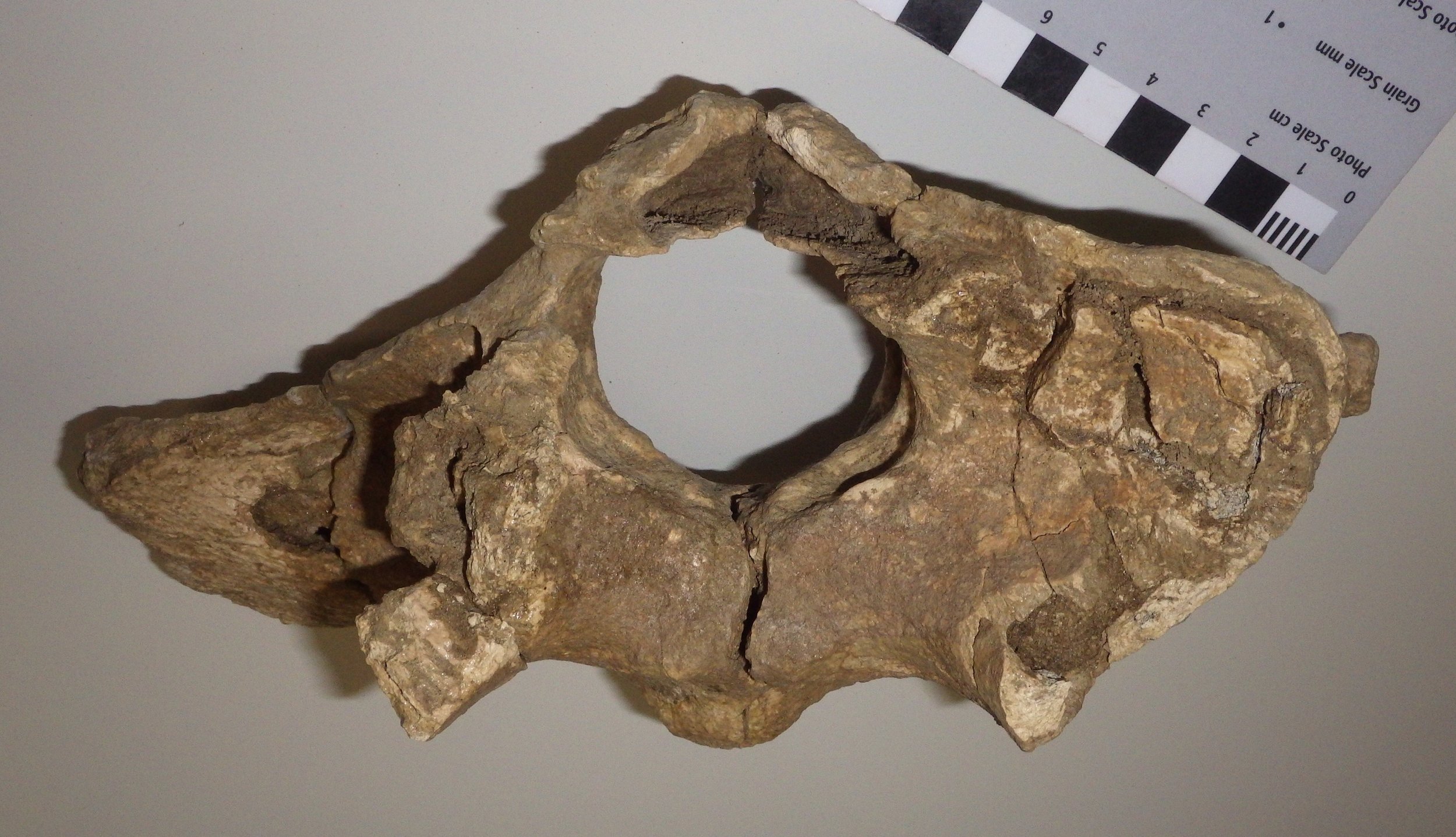
 For this week's Fossil Friday I'm going to stick with bison, this time featuring an atlas vertebra.As I've described in earlier posts, the mammalian vertebral column is generally divided into five different regions. The most anterior are the cervical (neck) vertebrae. Almost all mammals have exactly seven cervical vertebrae, but mammals are unique among vertebrate classes in having such a tightly constrained cervical count. The first two cervical vertebrae, the atlas and the axis, are always highly specialized and look very different from any of the other vertebrae.That is mainly because of the complex articulations necessary to allow a range of movement in the head. When you nod your head (flexion and extension), that motion primarily occurs at the joint between the front of the atlas vertebra and the skull. When you shake your head (rotation) the motion is mostly at the joint between the back of the atlas vertebra and the front of the axis vertebra. That all means that the atlas has to accommodate one type of motion (nodding) on its front surface, a different motion (rotating) on its back surface, while providing attachment points for all the muscles that actually cause the movement, and allowing a passageway for the spinal cord and the vertebral arteries and veins through the middle of all of this movement. It's no wonder that the atlas has such a distinctive appearance. (As an interesting aside, whales, which have almost no ability to move their heads, still have complex and unique atlas and axis vertebrae, a relict of their ancestors that had much more mobile necks.)So, with all that said, the image at top shows a bison atlas vertebra in anterior (cranial) view. The bone is somewhat crushed and incomplete; much of the right side in particular is missing (remember, since this is anterior view, the right side of the vertebra is on the left side of the photo). The large hole in the bone is the neural canal, the passageway for the spinal cord. Below and beside the neural canal (centered at the "5-o'clock" and "7-o'clock" positions) are large depressions that articulate with the occipital condyles on the skull; this is the main "nodding" articulation.
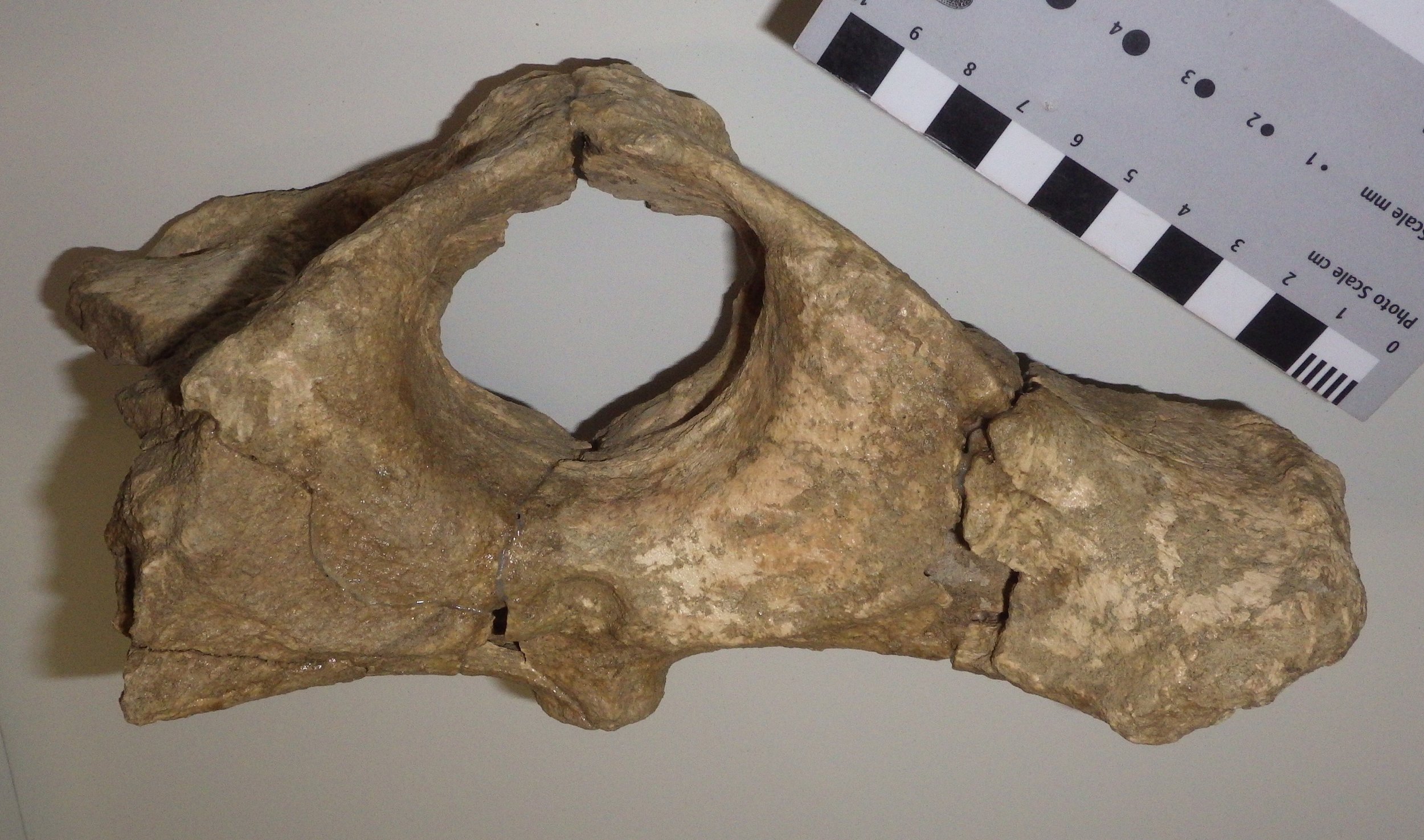 Looking at the same vertebra in posterior (caudal) view, we see broad, flat surfaces below and beside the neural canal. These are the articular surfaces for the axis vertebra, where rotation occurs. The large projection of bone on the right is a lateral or transverse process (the corresponding one on the left side is broken off). This provides a surface for the attachment of neck muscles associated with head movement. Bison have large, heavy heads, and so the muscle attachment areas to support them are relatively large.Here's the atlas in dorsal (top) view:
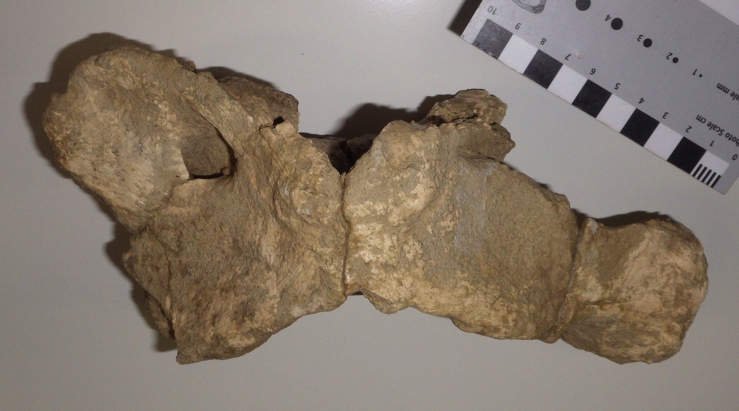 At this angle you can see that the transverse process is broad across the top surface, and has quite a large surface area. The opening toward the upper left is the alar foramen (the right one is only partially preserved), which provides a passageway for the vertebral arteries and veins.Even allowing for damage to the bone, the shape of this atlas seems a bit different from that of the modern Bison bison (see this site for an example). There are two different species of Bison known from the Diamond Valley Lake fauna, B. antiquus and B. latifrons. B. latifrons has a larger, heavier head than B. antiquus, and it's possible that this might be reflected in the anatomy of the cervical vertebrae, but I've not yet checked references to confirm this. At the moment, we have this particular atlas identified as Bison sp.