
 This week's Fossil Friday features one of Diamond Valley Lake's most common large mammals, the bison.Specifically, this bone is the atlas vertebra, the first vertebra in the neck. It's shown above in anterior view. The large hole in the bone is the neural canal, which carries the spinal cord in the live animal. The large concavities on each side of the neural canal are the articulations for the skull's occipital condyles.Below is the posterior view:
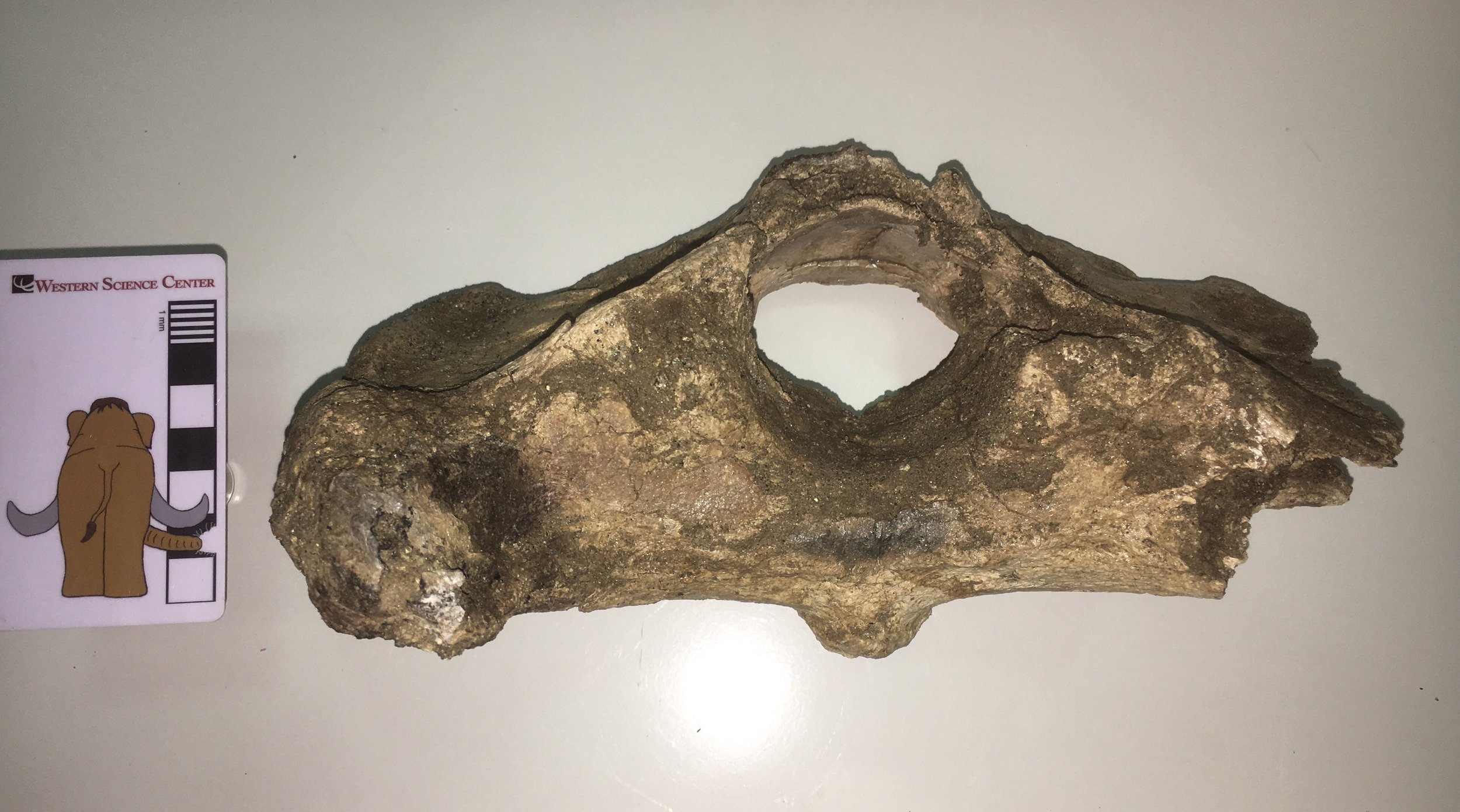 The preservation is a little rougher on this side. The posterior edge of the neural arch (the top of the neural canal) is missing, as is most of the right transverse process. The flat areas to each side of and slightly below the neural canal are the articular surfaces for the 2nd cervical vertebra, the axis.Here is the dorsal view:
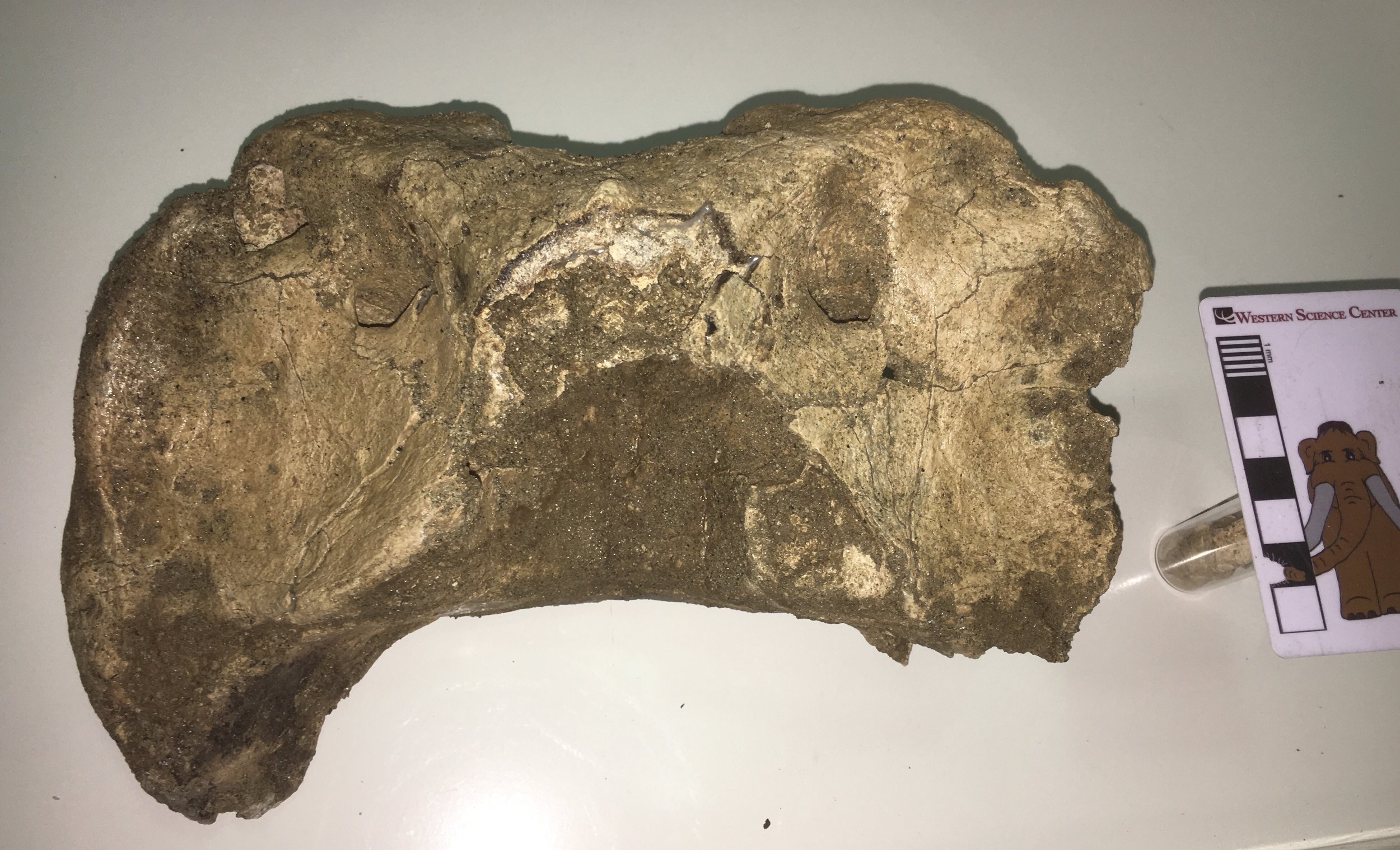 ...and the ventral view:
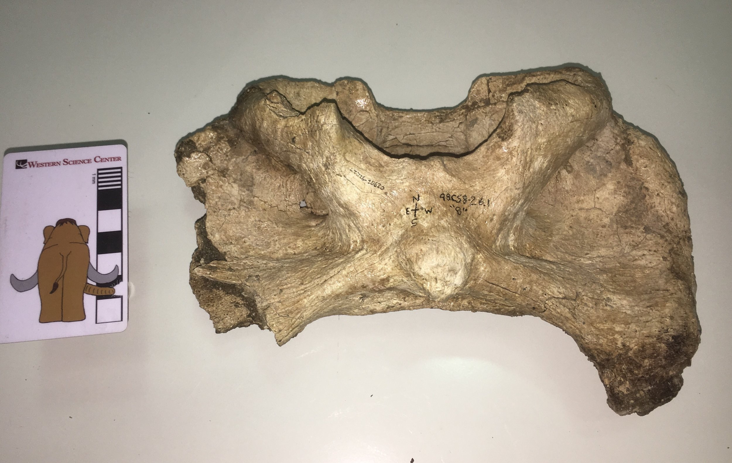 This vertebra is identified in our records as Bison antiquus. It comes from the West Dam area of the lake; sediments there are mostly less than 20,000 years old and seem to only produce Bison antiquus and not the large Bison latifrons. Bison antiquus is generally considered to be closely related or ancestral to the modern Bison bison, and indeed this atlas looks very similar to a modern bison's. This make an interesting contrast to the DVL bison atlas I wrote about back in May 2015. That vertebra was identified as Bison sp., but its shape is a bit different from this one (I even commented at the time that it differed from Bison bison). So perhaps our two extinct bison species really do have distinct atlas shapes. At some point we'll have to compare these bones to atlases that are associated with skulls to confirm that we're seeing specific differences and not individual or sexual variation.