 An oddity in the preservation of Max the mastodon is that, while the skull and pelvis were beautifully preserved, very little was recovered from the massive limb bones. The only significant piece was the distal end of the left femur, and a few months ago while working on our mastodon project we opened our exhibit case to photograph and measure this fragment.The image above is in anterior view. The smooth curved surface at the bottom of the bone is part of the articulation for the knee joint. Below is the same fragment in posterior view:
 One of the reasons we wanted to get measurements of this bone is that we've often stated (following Springer et al., 2009) that Max is one of the biggest mastodons known from the western United States. The femur is one of the better bones for estimating body size, because in most terrestrial animals it's the primary weight-bearing bone, so its dimensions are often indicative of both height and weight. Ideally, we would like to have the entire femur to measure, but we take what we can get. In Max's case we can get a good measurement of the maximum distal width, which gives us a point of comparison to other mastodons. The graph below shows how Max's femoral width compares to mastodons from Rancho La Brea and New York:
 The two left bars are Rancho La Brea specimens, the tall bar in the middle is Max, and the two bars on the right are respectively the Watkins Glen mastodon (an adult male) and North Java mastodon (an adult female) from New York (measurements from Hodgson et al., 2008). Max's femur is quite wide compared to other mastodons, which was part of the reason Springer et al. suggested that Max was such a large animal.Using these measurements, I made a sketch estimating the total size of Max's femur and compared it to images of the same four specimens from the graph, with the caveat that proportions often don't scale exactly linearly so this is only an approximation. In addition, since Max's femur is the left one and the other four specimens are right femora, I've reversed Max's image so they're more directly comparable. (The Watkins Glen and North Java images are from Hodgson et al., 2008):
 Any way you look at him, Max seems to have been a pretty big mastodon. Yet Max's teeth are relatively small. This disparity in Max's measurements is what started us looking at the tooth shape and size in California mastodons, and is something I'll be exploring further next month as part of our mastodon project.References:Hodgson, J., W. D. Allmon, P. L. Nester, J. M. Sherpa, and J. J. Chiment, 2008. Comparative osteology of Late Pleistocene mammoth and mastodon remains from the Watkins Glen Site, Chemung County, New York. In W. D. Allmon and P. L. Nester, eds., Mastodon Paleobiology, Taphonomy, and Paleoenvironment in the Late Pleistocene of New York State: Studies on the Hyde Park, Chemung, and North Java Sites. Palaeontographica Americana 61:301-367.Springer, K., E. Scott, C. Sagebiel, and L. K. Murray, 2009. The Diamond Valley Lake Local Fauna: Late Pleistocene vertebrates from inland Southern California. In L. B. Albright, ed., Papers on Geology, Vertebrate Paleontology, and Biostratigraphy in Honor of Michael O. Woodburne. Museum of Northern Arizona Bulletin 65:217-235.
Fossil Friday-mastodon jaw fragments
 We're entering the final hours of our crowdfunding campaign at experiment.com, and are very close to our funding goal. This week we're featuring another specimen that will be included in this study.While the bulk of the Western Science Center's collection came from Diamond Valley Lake, we are a regional repository with a number of collections from other localities. This specimen was recovered during a mitigation project in Temecula in southwestern Riverside County, and after reconstruction by WSC volunteers it became apparent we had a significant portion of a mastodon lower jaw. The image is in dorsal view, with anterior to the left. Parts of both dentaries are preserved, although there is considerably more of the left dentary present.Portions of four teeth are preserved, the lower second and third molars. On the left side the second molar is complete, and the third molar is nearly so, allowing us to get length and width measurements for inclusion in our project; it turns out that these teeth are long and narrow, exactly what we've come to expect from California mastodons. The second molars are pretty heavily worn, while the third molars show wear only on the first two lophs. That is very close to the same wear state found in Max, so these two were probably close to the same age.If you haven't already done so, please go to experiment.com/mastodon and donate to our project, so we can start to understand how mastodons were distributed through North America.
Fossil Friday - Old Man Mastodon
 Closing our our Month of Mastodons is one of the more complete mastodon skulls from Diamond Valley Lake, a specimen that has several interesting features in addition to its relatively good preservation.First, to explain what you're looking at, here's an annotated version:
 The skull is laying on its right side, at a slight angle. Much of the braincase seems to be missing, although this area hasn't been fully prepared, so there could be more there than there appears. The left dentary (lower jaw) is present, but disarticulated; it's not clear if the right dentary is still in the bottom of the jacket.So, what can we say about this skull? Well, for starters it looks pretty large, with a massive tusk. I haven't measured it out yet, but I suspect that based on size this will probably be a male mastodon. Zooming in on the teeth gives us more information:
 Both upper 3rd molars are visible, as is the lower left 3rd molar. These teeth are in wear across their entire occlusal surfaces. Also notice the flat area outlined in red below, located in front of the upper left 3rd molar:
 This is the closed-up socket for the 2nd molar, which has completely worn down and fallen out. With the 2nd molars gone and the 3rd molars heavily worn, this was a pretty old mastodon, most likely over 50 years old. He was much older than Max, who still had his 2nd molars, and in fact is the oldest mastodon I've seen from Diamond Valley Lake so far.There is another interesting feature in these teeth, specifically in the lower 3rd molar. The red line below traces the outside edge of the chewing surface of the tooth (the "labial edge of the occlusal surface"):
 The tooth is most heavily worn in the middle, and is higher both in the front and the back. This is the only time I've seen this wear pattern in a mastodon tooth, and I'm not sure how it formed. Here's the issue: like other advanced proboscideans, mastodons replaced their teeth horizontally. That means the front of the tooth erupts first. Because of this, at any given point in time after eruption, the more anterior parts of the tooth have been functional for longer than the more posterior parts. Because of this there should be a wear gradient, with the tooth being progressively less worn as you move from the front of the tooth to the back. And normally that's exactly what we see in mastodons, mammoths, and other proboscideans with horizontal tooth replacement. So how did this tooth get most heavily worn in the middle? Was the tooth injured or deformed in some way? Did the mastodon's diet change at some point, changing the wear rate? Did the mastodon suffer an injury to his jaw or jaw musculature that affected his ability to chew, altering the occlusion pattern of his teeth? (Max had multiple injuries to his lower jaw, so this type of injury may be common in male mastodons.) Or is there something else going on?This mastodon skull is currently on display in the "Stories from Bones" exhibit at WSC, but if you haven't seen it yet you'll have to hurry; "Stories from Bones" closes on May 29.With complete 3rd molars, this mastodon is also one of the specimens that is contributing data to our mastodon tooth research project, "Mastodons of Unusual Size". We only have a few days left in our crowdfunding campaign to support this research, and we're still well short of our goal, so donate today and help us understand mastodons a little better!
Fossil Friday - partial mastodon skull
 As we continue our month of mastodons in support of our crowdfunding campaign at experiment.com, this week we have a partial mastodon skull from Diamond Valley Lake.When I say partial, I mean very partial. Practically all that's left of this specimen are the upper teeth and one tusk. Almost all the bone is missing, a somewhat odd preservation type that we get occasionally at Diamond Valley Lake. We haven't taken measurements on the tusk, but it seems to be massive, suggesting that this might be a male. The skull is laying upside down, and oriented so that anterior is toward the camera (this large jacket was sitting on a storage shelf, and would have been difficult to move quickly to get Fossil Friday photos in other orientations). The two upper third molars are visible, but there's no preserved trace of the second molars. The crowns of the third molars are well preserved, so this is a specimen we'll be able to include in our study. Every loph on the molars is in wear, with the last lophs just starting to wear, so this animal was presumably a bit older than Max (who still had second molars and unworn last lophs on the the third molars).We have just under two weeks to go in our crowdfunding campaign to gather data on mastodon tooth shape from localities across the country, and we still have a long way to go, so please visit experiment.com/mastodon and give what you can to help us with this study.
Fossil Friday - skull of "Little Stevie" the mastodon
 As promised, during our crowdfunding campaign to work on mastodons I'm featuring mastodons in all my Fossil Friday posts. This week is the partial skull of "Little Stevie" from the WSC collections. "Little Stevie" was the most complete mastodon found at Diamond Valley Lake, with about 40% of the skeleton recovered. Thanks to a donation by Eric and Gisela Gosch, most of "Little Stevie's" skeleton is on display in a case embedded in the floor of the museum, but the partially prepared skull is in the collections. Below is a marked-up version of the image above:
 The skull is laying upside down, and the yellow arrow is pointing anteriorly. That means you're looking at the right side of the skull. The 2nd and 3rd upper molars are indicated in blue, and the right occipital condyle is labeled (the skull's articulation with the first neck vertebra). The broken cheek bones are visible close to the camera, as are the internal choanae (the internal openings for the nostrils) just behind the 3rd molar. There are also a number of postcranial bones in the jacket, including several ribs (there's one below the yellow arrow) and two thoracic vertebrae indicated by the red arrows. Both thoracic vertebrae are missing their epiphyses, indicating that "Little Stevie" was still growing. However, the femur associated with this skeleton has fused epiphyses, and at least the first two lophs on the third molar are in wear, which suggests that "Little Stevie" was close to full grown, maybe early to mid-20s.We don't know for sure if "Little Stevie" is a male or a female. The best markers for determining sex in a mastodon are the pelvis and the tusks, neither of which is well preserved in "Little Stevie" (although we may eventually be able to get a pelvic measurement). "Little Stevie's" femur is very close to the size and proportions of the Java Site mastodon from New York, which is thought to be a female, but that's not a very reliable indicator of sex."Little Stevie" does include both 2nd and 3rd molars and a femur, all of which are bones we're examining in our mastodon study, so that means "Little Stevie" is included in our project. Help us collect more data to compare to "Little Stevie" by donating at experiment.com/mastodon.
Fossil Friday - mastodon molar
 On Monday Eric Scott and I launched a crowdfunding campaign to support a mastodon research project we've been working on. We believe California (or maybe western) mastodons have different tooth proportions than mastodons from other part of the country, and in order to test that hypothesis we need to travel to other museums to collect measurements on mastodon specimens from as many locations as possible. In recognition of that campaign, for the next month all my Fossil Friday posts will feature mastodons.Above is a left upper 3rd molar from Diamond Valley Lake, shown in occlusal view. This tooth was found shattered and painstakingly reconstructed, so even though it's incomplete it's an impressive specimen. Below is a lingual view of the same tooth:
 The anterior loph (the large ridge on the crown of the tooth) is mostly missing on this specimen, but the other lophs show little or no wear, so this tooth was either unerupted or had only recently erupted. As an aside, while I haven't checked the numbers to confirm this, a lot of the Diamond Valley Lake mastodons seem to have died when the third molars were partially erupted, maybe in their mid-20s to mid-30s.Since the front of this tooth is missing we can't take a reliable measurement of its length, which means this specimen will probably not end up in our project database; we need to be able to compare the length and width of the tooth crown. But even incomplete, this seems to be a remarkably small tooth even by California standards. Interestingly, our data so far suggests that molar size is not a very good indicator of body size (Max was a big animal with relatively small molars), so even though this tooth is small it doesn't mean it came from a small mastodon.We hope you'll visit experiment.com to help us study these mastodons!
Fossil Friday - mastodon thoracic vertebra
 With over 100,000 Pleistocene fossils in the WSC collections, there are still many that I've never seen. There are others that I've glanced at dozens of times without ever taking a close look, like the mastodon vertebra shown here. As a result, interesting stories are often in plain sight, unnoticed until someone takes a close look.The bone is shown above in anterior view, and is a thoracic vertebra based on the rib articulation visible on the right side of the image. It appears to be a fairly posterior thoracic, although I haven't tried to identify the exact position. The vertebral centrum is heavily eroded, especially on the ventral side, but the dorsal part is better preserved. This vertebra was associated with a few scrappy remains of at least one other vertebra, but that was all that was recovered from this individual.In dorsal view we can see the base of the neural spine and the top of the neural canal. This is where things get a little strange:
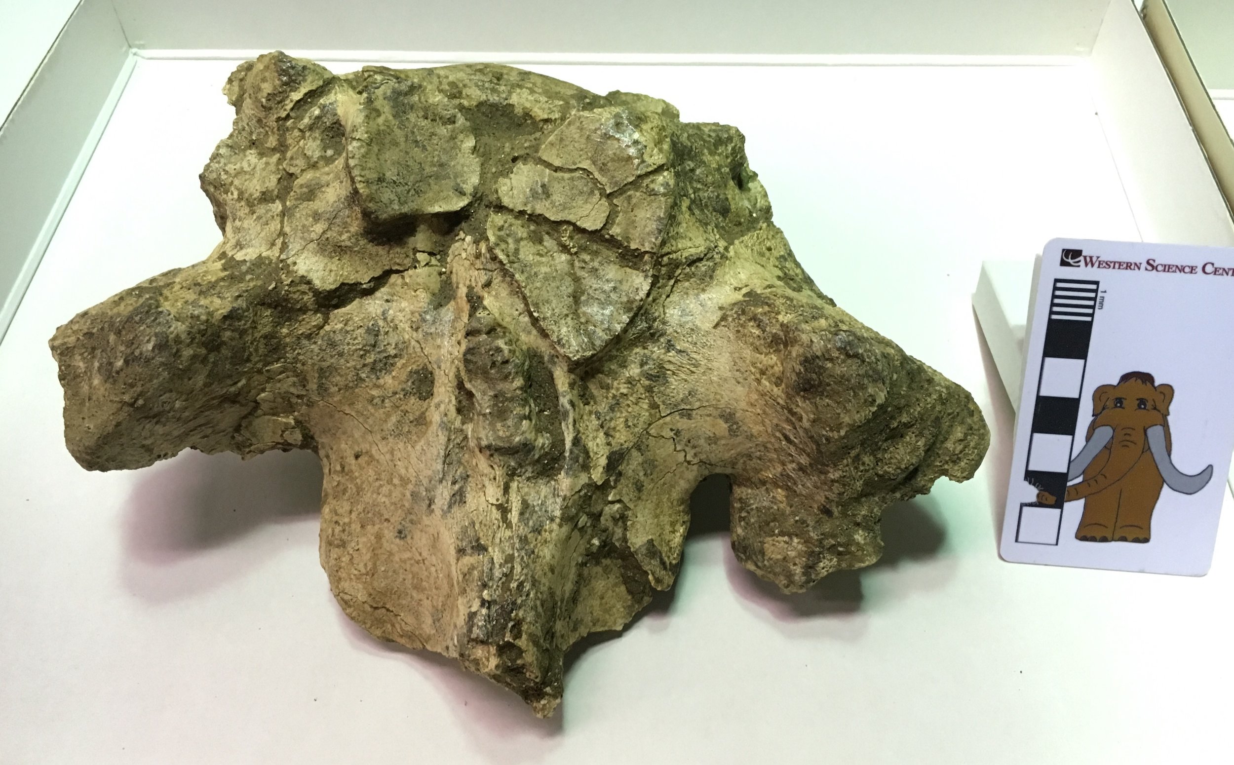 Typically, a vertebra articulates at three points with each vertebra in front of and behind it. Two of those points are the anterior and posterior surfaces of the centrum (so the anterior surface of the centrum articulates with the posterior surface of the centrum in front). The other articulations are located on the neural arch or spine. These articulations are often found at the tips of projections called zygapophyses, so the two prezygapophyses on the front edge of the neural arch will articulate with the corresponding pair of postzygapophyses on the next vertebra in front. The actual anterior articulation areas are called the prezygapophyseal articular facets (because zygapophysis was not an intimidating enough term by itself)!In mastodon thoracic vertebrae the prezygapophysis doesn't really stick out as a projection, but the articular facets themselves are very large. Below is the dorsal view image with the articular facets outlined in red:
 The left and right prezygapophyseal articular facets should be nearly mirror images of each other, but that's clearly not the case here. The right facet is more than twice as large as the left, and is actually so large that it extends slightly across the midline of the vertebra onto the left side. In addition, note the area outlined in blue, shown below in an oblique closeup:
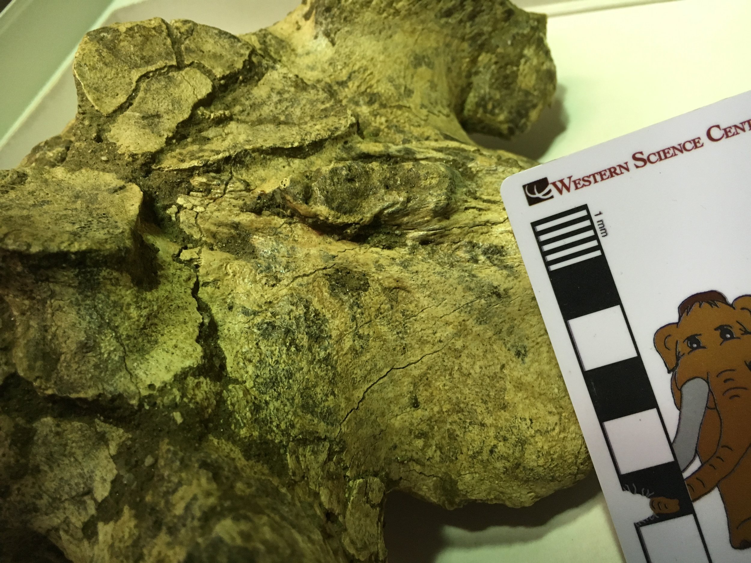 This is an osteophyte, a pathological bony growth that may form due to injury or as a result of osteoarthritis. It's not clear exactly what happened to this mastodon. The vertebra could have grown abnormally due to a genetic condition, and the resulting unusual stress caused by a deformed vertebra could have caused osteoarthritis later in life. Alternatively, the mastodon could have suffered an infection or injury that resulted in the deformed vertebra. The injury needn't have occurred in this spot; a muscle or bone injury elsewhere in the back or even in the legs or feet could have placed enough asymmetrical stress on the back to cause this deformation. Basically, walking with a limp over a long period of time could have potentially caused a condition like this. Since we have only a few bones from this animal, it's likely we'll never know for sure why this mastodon had such an asymmetrical vertebra.
Fossil Friday - mastodon palate
 Taphonomy is the subset of paleontology that examines how an organism gets preserved as a fossil. Taphonomists consider all kinds of different features, including the structure and composition of the organism, how and where it died, rates of sedimentation, the quantity and chemical composition of groundwater in the area, and countless other variables. Understanding taphonomy can tell us a great deal about an organism and its environment, and it's something to be considered when looking at unusual preservation.The specimen shown here is the partial palate of a mastodon, seen in palatal view (looking up at the roof of the mouth). All that remains are the crowns of the teeth and few bits of the maxillary and palatine bones. I'm not sure why this specimen preserved this way, but there are some possibilities to consider. By far the best preserved portions are the crowns of the teeth. Tooth enamel is generally the hardest and most chemically resistant part of the vertebrate body, so it's not entirely surprising to see teeth surviving while the rest of the skeleton deteriorates.This specimen has both left and right teeth preserved, in their correct life positions relative to each other, even though most of the bone between them is gone. That means the upper jaw survived as a unit for at least some period of time. Perhaps the skull was sitting on the ground in such a way that the upper jaw was buried but the rest of the skull was exposed and eroded away. Maybe when the animal was rotting or scavenged, the maxillae separated from the rest of the skull. Or maybe the skull was largely intact when it was buried but groundwater mostly dissolved the bones and left the teeth behind (we have a horse in the collection that may have experienced this).As for the mastodon itself, the specimen is oriented with anterior to the left, so the left teeth are at the top and the right teeth are at the bottom. Portions of the crowns of both upper 2nd molars are preserved, and the left one is almost complete. The crowns of both upper 3rd molars are also preserved. The 2nd molars show heavy wear, but the 3rd molars show very little wear, and only on the anterior part of the tooth. This suggests a mastodon that was probably in its early 20s when it died.---As a reminder, tomorrow is the 2nd Annual Inland Empire Science Festival at the Western Science Center. Come to the museum to for tons of science-themed activities, lectures, and exhibits. Student, youth, and under-4 admissions will receive a coupon for a free replica Tyrannosaurus tooth, molded from a specimen on exhibit at the museum.
Fossil Friday - mastodon pelvis
 When the permanent exhibits were being installed at WSC, several high-profile specimens were molded and cast for the displays, including the large mastodon nicknamed Max. It turned out that Max's pelvis was too fragile to withstand molding, so while the pelvis is on exhibit it was the only recovered element of Max's skeleton that wasn't reproduced.The mammalian pelvic girdle is a complex structure made up of multiple bones that are generally separate in young animals but mostly fuse with age. All of the various pelvic bones are preserved in Max, although some of them are incomplete.The image above shows the pelvis in close to dorsal view, maybe somewhat posterodorsal (above and behind), with a Max scale bar for scale. It is rotated so that anterior is toward the top of the image. Below is a marked-up version of the same photo:
 The largest bone, outlined in blue, is the ilium (only the bones on the right side and the midline have been color-coded). The ischium is outlined in red, and the pubis, partially hidden under the overlying bones, is yellow. The bulge on the side where the ilium and ischium meet is the hip socket, or acetabulum. The actual socket opens to the side and down, so it's hidden in this view. The pubis also comes up to the bottom of the acetabulum, but that is also hidden in dorsal view.The left and right ilia are separated by, and fused to, a structure called the sacrum, which is made up of a series of fused sacral vertebrae, outlined in purple above. Max appears to have four sacral vertebrae; the number can vary on an individual basis, and some mastodons have five. The first two tail vertebrae (caudal vertebrae) are also present, outlined in dark blue. It's not clear if they are actually fused to the sacrum, or simply interlocked with it. Excluding the tail vertebrae, Max's pelvis is therefore made up of 10 bones: the left and right ilia, left and right ischia, left and right pubes, and four sacral vertebrae.Besides serving as the attachment points connecting the legs to the body, the pelvic girdle supports the back of the abdominal cavity. The digestive tract exits the abdominal cavity through the urethra and anus, so the pelvic bones are arranged in a tube to allow for this, as well providing a path for the birth canal in females. The sacrum forms the top of this tube, while the ilia and ischia make up the sides and pubes make up the bottom. This is more obvious is we look at the pelvis from below and behind (posteroventrally) - exactly where you don't want to stand with an elephant that has recently eaten or that's about to give birth:
 Below are two more views of Max's pelvis, to give a better idea of the arrangement of the various bones. First, an oblique view from above the right side (anterior is to the lower right):
 And above the left side (anterior is to the lower left):
 Notice in each of these views, and especially in the marked-up image, that the boundaries between the various bones are not immediately obvious. Max, while not elderly, was a fully mature mastodon (probably over 30 years old), so these bones were solidly fused to each other. In a younger mastodon the boundaries between the bones would be more obvious.Max's pelvis only has a partial support cradle, so unfortunately we can't flip it over to see the ventral side. Even so, we can see quite a lot in the views that are available.
Fossil Friday - mastodon rib
 Here at Valley of the Mastodon, we're ringing in the new year with a rib from our namesake animal. Ribs don't often get a lot of attention, but there sometimes is information that can be gleaned from them.The left rib shown above is incomplete, with portions of both ends of the bone missing. The proximal end, closest to the vertebrae, is to the right. This rib is large enough to be from an adult or near-adult animal, but it's impossible to estimate the age with any certainty. Ribs do have an epiphysis at the proximal end that is unfused in young animals, but that area's not preserved in this specimen.So what's going on with the proximal end of this rib? Here are some closeups from various angles:

 A swollen region around the bone, pits and bone spurs on the surface...these are all indications of an injury or other pathology. As with many of the specimens in our current "Stories from Bones" exhibit this bone shows a healing response to some type of trauma. It's difficult to say what the trauma was. The bone could have been broken in an attack by a predator, and while there are no indication of bite marks to corroborate this it's still a possibility. The rib could have been broken in a fall or some other accident, or in a fight with another mastodon. Infections and certain diseases can also sometimes cause this type of response in a bone, but we are definitely looking at something that happened to this mastodon while it was still alive.
Fossil Friday - mastodon molar
 I've been looking at a lot of mastodons lately, so they're likely to show up more and more on Fossil Friday. Today's entry is an upper left 1st molar, collected from the West Dam at Diamond Valley Lake.The tooth is shown above in labial view (the side of the tooth on the outside of the mouth, literally closest to the lips), with anterior to the right. Of course, since it's an upper tooth, I really should have photographed it with the crown at the bottom.Here's the lingual side (literally, closest to the tongue):
 Notice that the pointed parts of the crown (called lophs) are more worn in this view. In mastodons (and in many other mammals with grinding molars) the crowns of upper teeth will wear more rapidly on the more the labial side. The reverse is generally true of the lower teeth.Here's the occlusal view of the same tooth (with anterior toward the bottom):
 While the labial side may be somewhat more worn, in this view we can see that there's heavy wear across the entire surface. Even so, had this mastodon lived longer, this tooth could have remained functional for quite some time. There roots are intact, and there's still a lot of enamel all the way around the margins of the tooth that had not yet worn away.
Fossil Friday - mastodon cervical vertebra
 Elephants and their relatives have big heads. They're big animals to begin with, but they also have strong, heavy jaws and teeth, as well as big brains. Then they throw in a massive trunk and huge heavy tusks (four of them, in the case of some extinct species). If you stick all these heavy features onto your skull, there are going to be consequences.Two things we very rarely see together in one animal are long necks and large heads. In some ways the head and neck act as a third-class lever, which actually increases the force necessary to move the head. The longer the neck the greater the necessary force. Significantly, that also applies to the force needed to stop the head from moving once it's started; without enough force, the head may keep moving independent of the neck and the rest of the body, with unfortunate consequences. What this boils down to is that animals with big, heavy heads will almost always have short necks.The image at the top is the third cervical (neck) vertebra from a mastodon, seen in anterior view, and below is the same vertebra in posterior view:
 The vertebra has both epiphyses attached, which suggests that it was not a particularly young animal (we can't say that it was fully mature, since the cervical epiphyses fuse at a relatively young age). There are some hints of osteoarthritis. This could indicate that the animal is older, but I wonder if this is common in elephant neck vertebrae, given all the stresses on the neck.The right lateral view is especially interesting:
 This vertebra is amazingly short. If I had found it in a marine deposit I would have probably initially thought it came from a whale - another large-headed animal with short neck vertebrae.In mastodons and other proboscideans, the third through seventh cervical vertebrae are all short like this. A string of these short vertebrae is not going to be very flexible, so one of the trade-offs in having a big, heavy head is having a short, inflexible neck. In most whales and dolphins some of the neck vertebrae are actually fused together making them completely immobile. This hasn't happened in elephants, but they still have very limited neck mobility. As an example, check out videos such as this one in which elephants are facing off with possible threats. Notice that as the elephant in the video looks in different directions, he's actually turning his whole body. His head is staying in almost the same position relative to his back the whole time. Elephants can still nod their heads and rotate their heads around the neck, because these motions are largely controlled by the first two neck vertebrae. But they have very little ability to point their head in a different direction than the rest of the vertebral column, a limitation that they share with their mammoth and mastodon relatives.
Fossil Friday - juvenile mastodon humerus
 It's been over two months since we've featured a mastodon for Fossil Friday, which seems a little odd for the Valley of the Mastodons, so this week we have a mastodon humerus.This is a left humerus (the upper arm bone), seen above in approximately anterior view. The proximal end (closest to the shoulder) is on the left, while the distal end (closest to the elbow) is on the right. Below is the same bone in posterior view:
 The ends of the bone are indistinct and have no obvious articulations with other bones. While there is some damage to the bone, the primary reason for this is that this was a very young mastodon. The humerus starts out as three different bony components, a main shaft and epiphyses at each end that are all held together by cartilage. As the animal grows the elements eventually fuse together into a single unit, but if the animal dies before it's fully grown the epiphyses may fall off as the cartilage decays. That's what's happened in this case, with both the missing epiphyses and the small size indicating that this was a very young mastodon.
Fossil Friday - Stories from Bones exhibit
 For Fossil Friday this week, I want to highlight Western Science Center's new exhibit "Stories from Bones", which opens tomorrow.While WSC has excellent paleontology exhibits, as with any museum with a large collection many of the specimens are not on public display. There are a variety of reasons for this. Of course, the biggest obstacle is money; cases, information panels, interactive, floor space, and other requirements for an effective display are all expensive, and even the healthiest museums operate on a shoestring budget. Besides money issues, many specimens are just not suitable for display. Perhaps they're too fragile to risk moving around too much, or too fragmentary to interpret for the public (a specimen that visually looks like a piece of junk can still produce valuable scientific data). Even with all these limitations, we strive to make as much of our collections accessible to the public as possible. "Stories from Bones" is a result of that effort.
 Mammoth jaw display in "Stories from Bones".
An important aspect of planning an effective exhibit is developing a theme. An exhibit is telling a story, and you need to be aware of what that story is as the exhibit is being designed. The theme might be "We have a bunch of stuff!", but while that was a common theme in museums a century ago (and one I personally appreciate), it does not generally make for the most informative exhibit experience for the majority of visitors.Once the theme is established, it's important to stick to it, so that the exhibit story remains coherent. Imagine reading a mystery novel in which three chapters are devoted to a history of the development of the gunpowder used in the crime, and an additional chapter describes the etymology of the last name of the victim, when neither is important to the outcome of the story. Each of these things might be individually interesting, but if you try to talk about all of them then you risk obscuring everything. There is a real risk of this "mission creep" in an exhibit based on a data-rich field such as paleontology. We might talk about evolutionary relationships, paleoenvironmental indicators, biogeographic information, site-specific descriptions, or an array of other things. Talking about any of these might be a good idea; talking about all of them is a bad idea.The permanent paleontology exhibit at WSC does this very well. The exhibit is basically a review of the Diamond Valley Lake Local Fauna; what was here, how does it compare to the rest of Southern California, and (as a secondary point) what does it tell us about the local Pleistocene paleoenvironment. In contrast, "Stories from Bones" asks "What do these fossils tell us about the lives and deaths of these individual animals?".To that end, "Stories" has a series of displays that talk about how paleontologists determine how old an animal was when it died. We have several cases that look at tooth replacement in proboscideans, horses, and bison, such as the two mammoth jaws above (they're close to the same size, but one animal was about 30 years older than the other), or the three bison dentaries shown below that represent young, middle-aged, and elderly animals.
 Bison jaw display in "Stories from Bones".
We have several examples of bones that were broken and healed, evidence of events that took place during an animal's life:
 Broken and healed bones in "Stories from Bones".
We also have several cases that describe taphonomic features, looking at what happened to an animal at or immediately after death.We designed and built a number of interactive displays for this exhibit. The most prominent is a cast and video of the CT scans of Max the Mastodon's lower jaw, taken back in August.
 Max's CT-scan station in "Stories from Bones" during installation, under the watchful eye of @MaxMastodon.
We're proud of the fact that several of our interactive displays ask visitors to map or measure specimens and reach conclusions based on their data:
 A more extensive version of the bison tooth display shown here is also available as a guided activity for school groups visiting the museum, and as a kit available for purchase.If you're a regular reader of this blog, you'll find that "Stories from Bones" draws heavily from my past "Fossil Friday" posts. For most of those specimens, this is the first time they've ever been on public display, so if you're near Southern California make sure to stop by the museum. "Stories from Bones" opens on October 31, and will remain open into May 2016.
Fossil Friday - Max's mandible, CT-edition
 Last week we took Max to California Imaging and Diagnostics in Hemet, who donated a some time on their CT scanner to scan the lower jaw of Max the mastodon. A couple of days later CID provided us with disks with the scan data, and we've finally had a chance to start examining them. If you're unfamiliar with CT scans, the term is short for X-ray computed tomography. CT scans take a large number of X-ray images from multiple angles. A computer can then combine those images to make cross-section X-ray slices through the object. The slices can be stacked to make a digital 3D model of the object, or removed to examine the internal structure at any particular slice. It's an invaluable, non-destructive method of examining the inside of an object.I've uploaded an initial video of the slices of Max's jaw. The slices are transverse, run anterior to posterior, and are viewed facing anteriorly. Basically, imagine standing behind the jaw looking forward, and seeing cross-sections of the jaw gradually getting closer to you:http://www.youtube.com/watch?v=hK8c5pAgUrsSince the video runs front to back, the first thing you see is the anterior tip of the jaw. (Well, actually the the first thing is the plaster cradle the jaw is sitting on, then the tip of the jaw.) As you move back, you pass though the mandibular symphysis where the two haves of the jaw are joined, through the teeth, and then to the coronoid process and mandibular condyles (the condyles are incompletely shown, because the jaw was a bit to large for the scanning area).There are several slices that have especially noteworthy features. This is about 4 seconds into the video:
 The image of the jaw on the right is for orientation; the purple line is the approximate location of the slice. The blue arrow is pointing the pathological bone growth near the tip of the right dentary. Inside the bone, there are a series of fractures next to this bony growth, one of which is marked with the green arrow. I'm not sure if they are actually associated with the growth, or if they occurred after burial.Moving a little further back, to about 6 seconds:
 We're now behind the bony growth, but its effects are still present. The blue arrow is pointing to a foramen (a passage for nerves or blood vessels) that has been displaced anteriorly and ventrally, apparently in response to the growth. The green arrow is pointing to the mandibular symphysis, where the left and right dentaries are joined together. This was a surprise to me. These bones fuse together in older animals, obscuring the line of fusion. Max was a fully mature mastodon, middle aged at least. I would expect the symphysis to be completely fused, and on the outside it is; there's no trace of the symphysis on the outside of the bone. Yet inside they're still unfused. Is this typical of mastodons, or the Max's symphysis never fuse (or pop open) due to the beating his jaw apparently went through?Moving further back, to about 8 seconds:
 This slice shows how much that foramen was displaced, when compared to the left dentary (blue arrows). It's also clear that the dentary is still messed up well behind the bony growth; note how asymmetrical the two dentaries are on the dorsal surface (they should be nearly mirror images). In fact, the entire mandible is quite asymmetrical. I originally assumed that this was mostly due to deformation that occurred after burial, but it's now clear that at least some (maybe most) of the asymmetry occurred while Max was alive.At around 14 seconds:
 We're now getting into the tooth rows. The first root and cusp of the left 2nd molar is clearly visible (yellow arrow). The white arrow below the tooth is the opening of another foramen. One the right, the notch marked by the green arrow is the socket for the last root of the right 1st molar. Max was a mature mastodon, so the 1st molars (and all the premolars) had already worn down and fallen out; only the 2nd and 3rd molars remained, and the 2nd molars were heavily worn.As we already knew from examination of the exterior, Max had a second injury on his right dentary, essentially a crease running obliquely below the anterior tooth row with ridges of secondary bone growth on each side of the crease. This injury is marked with the blue arrow, showing apparently compressed bone beneath the surface (the bright line on the inside) and the swelling associated with the secondary bone growth.From this point in the video you can see the tooth cusps and roots grow and shrink as the scans run through the tooth rows. There is a change in one of the teeth at around 28 seconds:
 At this point we're all the way back to the last root of the 3rd left molar. As indicated by the green arrow, the pulp cavity on this tooth is open (you can see the same thing on the last root on the right 3rd molar at 29 seconds). This suggests that this part of the tooth was still growing. Sure enough, if we look at the cusps, the last cusp on the 3rd molar is only very slightly worn, indicating that it had just erupted.By 34 seconds the scans have passed completely through the tooth rows, and the remainder of the scans pass through the coronoid processes and the mandibular condyles.We still have a lot to look at with these scans, and we're hoping they'll be useful to people that are working on mastodon anatomy. We're going to keep working to make this data available online as we have time to process it. We also intend to include annotated versions of these scans in our upcoming exhibit, Stories from Bones.
Fossil Friday - Max the mastodon's mandible
 If you follow the museum's social media pages, you' re probably noticed that last Tuesday we had an adventure with one of the museum's mastodons, Max. We pulled Max's lower jaw off exhibit and, via ambulance and with a police escort, sent him to California Imaging and Diagnostics in Hemet for X-rays and CT-scans. There were several reasons behind this effort, but the primary one was that Max's jaw shows evidence of several injuries that we wanted to further explore.
 Max leaves the museum amid a crowd of Western Center Academy students.
 Preparing to load Max into the ambulance. One of Max's injuries is very obvious and we've known about it for some time. There is an anomalous bony growth at the right anterior tip of the jaw:
 The ridge anterior to the teeth on the dorsal side of the jaw is also rather misshapen in this area, possibly related to the same condition.The second injury was first spotted a few months ago, and was actually first observed in a cast we were making of the jaw; in the original specimen the injury was hidden due to the case structure and lighting the exhibit. This injury is a 2 cm-wide groove, running diagonally along the right dentary below the second molar. There are ridges of swollen secondary bone (a healing response to a bone injury) on each side of the groove:
 We're still processing the data gathered at CID, and I'll have more on that (including images) in a future post. But there are already some things we can say about these features, starting with the fact that they occurred while Max was still alive, at least weeks or months before his death(and possibly years before). That's because both feature show evidence of bony growth, and it takes time for that to occur. Evidence of healing is the most reliable method of determining whether a break in a fossil bone occurred before or after death.Max is a large mastodon, the largest reported from California. Given the size and the massive tusks it's probably that Max was a male mastodon. That, and the fact that he was a mature adult (based on tooth wear), suggests a possible cause of these injuries: intraspecies combat. Like modern elephants, there isabundant evidence that male mastodons engaged in intraspecies combat (see Fisher, 2009 for example), often resulting in injuries, sometimes fatal injuries. It's possible that both of these injuries, especially the groove on the side of the jaw, were caused by the tusks of another mastodon.Reference:Fisher, D. C. 2009. Paleobiology and extinction of proboscideans in the Great Lakes Region of North America. In G. Hayes (ed.), American Megafaunal Extinctions at the End of the Pleistocene, Springer Science: 55-75.
Fossil Friday - mastodon caudal vertebra
 Even in huge animals, not every bone is large. This small bone, less than 4 cm in length, comes from one of the largest Pleistocene animals at Diamond Valley Lake — a mastodon.This particular specimen is a caudal, or tail, vertebra. Its simple shape is not due to broken and missing pieces; in fact the bone is almost complete. The prominent spines and processes found sticking out on most vertebrae tend to be reduced or absent on caudal vertebrae in most animals. This is unsurprising from a functional standpoint. Those projecting spines are not simply decoration; they provide anchor points for muscles that control movement of the head, the legs, or other parts of the body. For the most part the tail doesn't serve as the anchor point for any muscles, and so the vertebrae don't generally have large processes. (There are a few exceptions: the first few caudal vertebrae have large processes that anchor the muscles that control the movement of the tail itself. There are also some animals such as alligators that have flattened tails, generally for swimming, which have enlarged caudal spines.) Their simple shape can sometimes make caudal vertebrae tricky to identify. They are sometimes mistaken for toe bones, but in most terrestrial animals even toe bones will have complex articulations and muscle attachment points that caudal vertebrae generally lack.The complete lack of processes indicates that this vertebra is from fairly close to the tip of the tail. To be that far back and still have a length of almost 4 cm indicates that it actually came from a big animal, and in fact other vertebrae associated with this bone indicate that it came from a mastodon.Looking at the bone end-on (below) we can see that the end is fairly rough:
 This is because the bone is missing the vertebral epiphyses, the caps of bone that fit onto each end of the vertebra. The epiphyses remain essentially as separate bones attached to the vertebra with cartilage while the animal is still growing, but eventually fuse onto the vertebra. The fusion of all the various epiphyses indicates that the animal has reached physical maturity; in elephants (and people) this is generally sometime around age 20-25. The lack of fused epiphyses in this vertebra (and, in fact, none of the epiphyses are fused and the other bones associated with this specimen) indicates that it came from a young animal, likely not more than a few years old.
3D models of WSC specimens
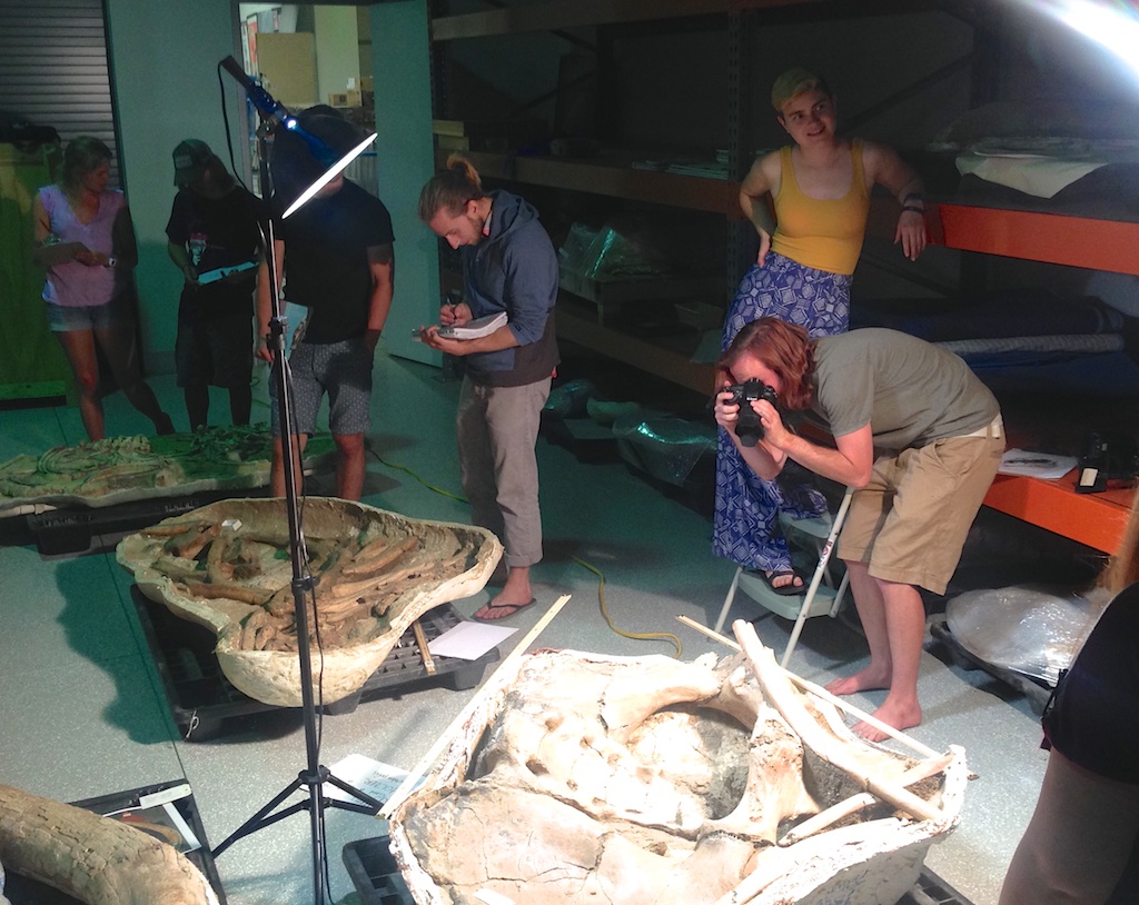 Last Saturday Pomona College's Karl Lang brought his geology class on a field trip to the Western Science Center, to learn about California's Pleistocene fauna and museum operations. While they were here, the students also took photos of several mastodon jackets in our collection in order to produce digital 3D models, which they've been kind enough to post online.Each image below has an embedded link to take you to a 3D viewer. Within the viewer, the images can be rotated and zoomed (I unfortunately couldn't get the viewer to embed properly in Wordpress).A partial vertebral column and ribcage:
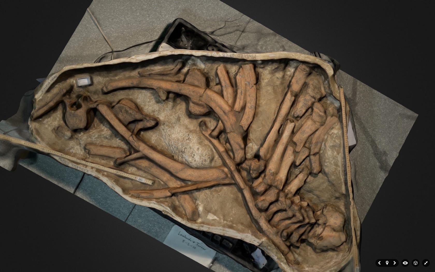 A pelvic girdle:
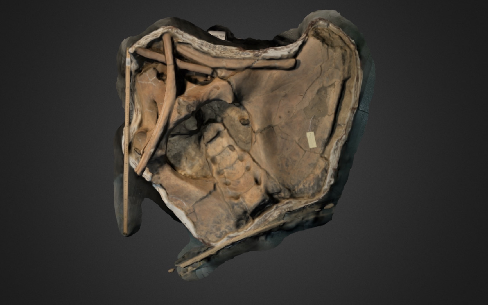 A partial skull:
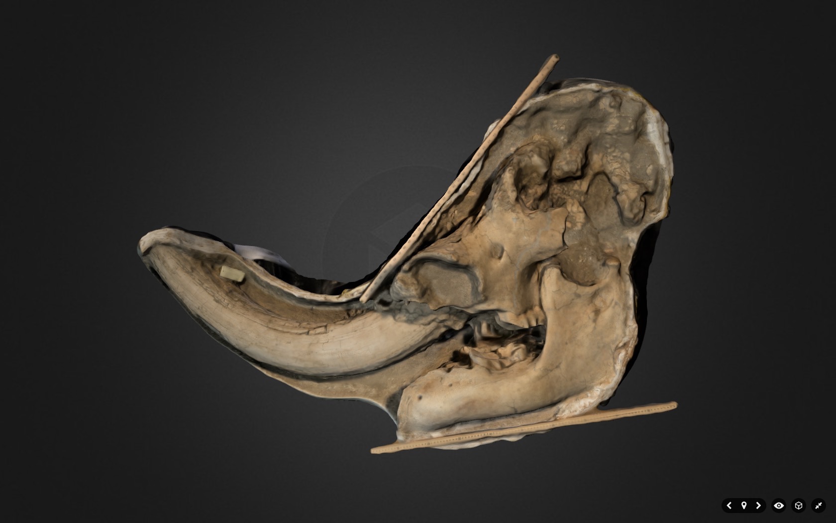 Thanks to Karl and Pamona's geology students for visiting, and for producing these great models!
Fossil Friday - juvenile mastodon femur
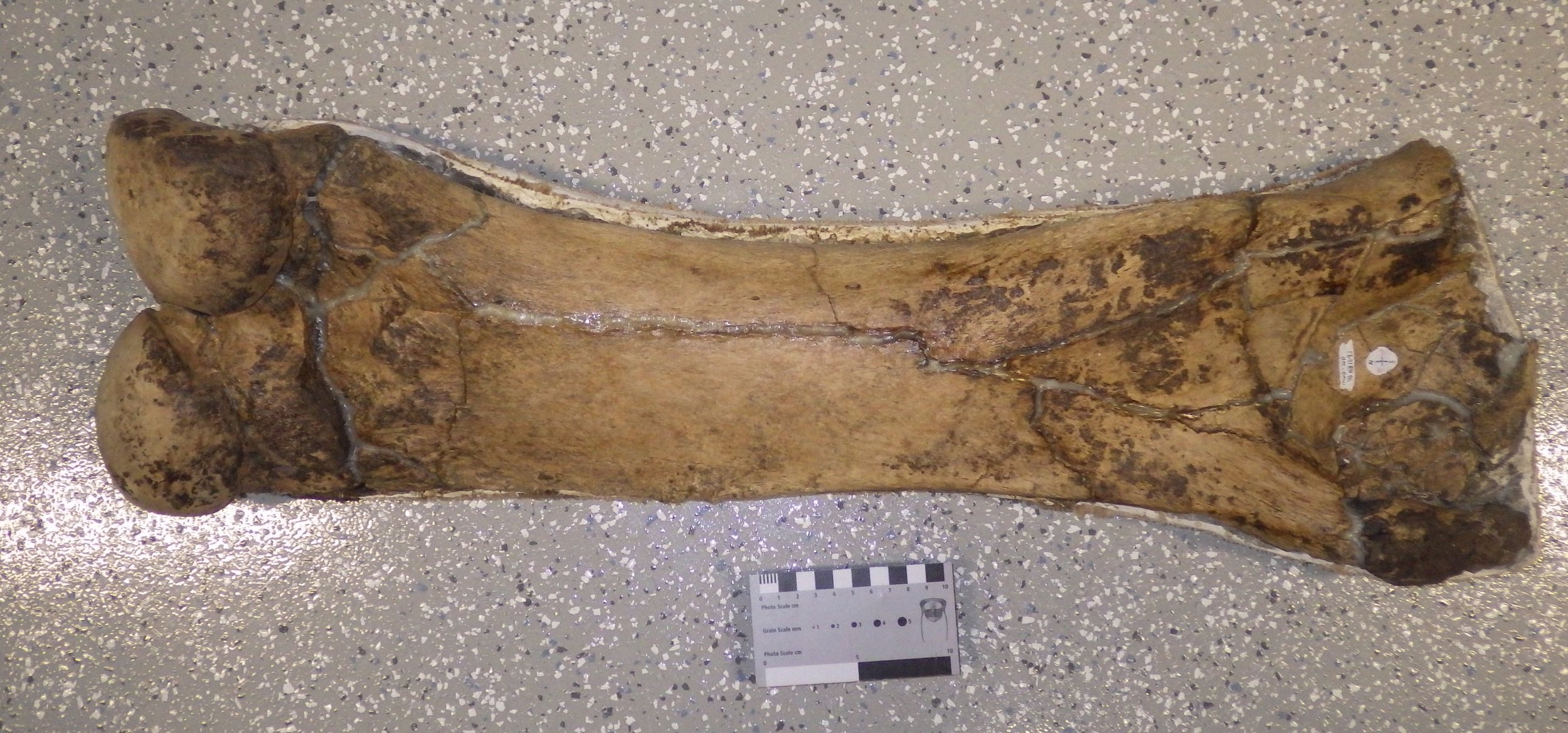 This week's Fossil Friday specimen is a femur (thigh bone) from a mastodon, collected from the West Dam of Diamond Valley Lake.Like many of the Western Science Center specimens, this femur is only partially prepared, and still sits in its field jacket. The posterior side of the bone is shown above, with the back of the knee joint visible on the left. As this is the right femur, there should be a large ball joint (the femoral head) in the upper right, that would articulate with the hip socket. Unfortunately the femoral head was broken off and not preserved.The preserved part of this bone is roughly 70 cm long. That's actually pretty small for a mastodon, large specimens of which can have femora more than a meter in length. This was probably a sub-adult mastodon, although the partial fusion of the knee joint to the rest of the bone suggests that it wasn't too young.
Fossil Friday - baby mastodon teeth
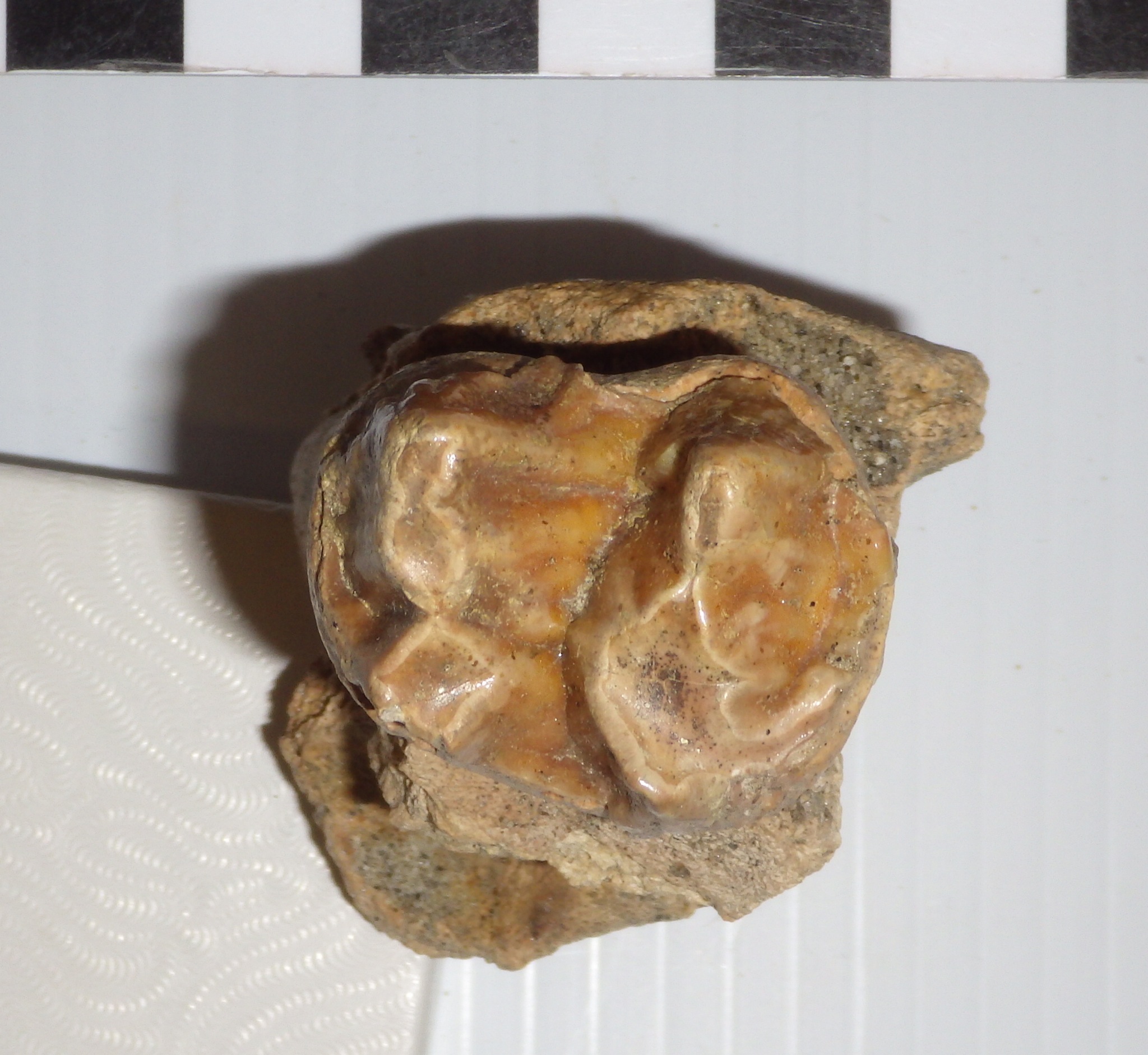 It seems that most of the mastodon remains from Diamond Valley Lake are from adult or nearly adult animals, although there are exceptions representing younger animals. Then we have the example shown here, from an almost ridiculously cute baby mastodon.This tooth is the upper right second premolar (dP2), the very first upper tooth to erupt in mastodons. It's shown above in occlusal view, with the front of the tooth to the left. Mastodon teeth have distinctive transverse ridges of enamel (they're what make tooth look "bumpy"), and the number of ridges differs by tooth position. Typically the second and third premolars each have two ridges, the fourth premolar and first two molars each have three, and the third molar has five. The two ridges in this tooth make it either a second or third premolar, but its size gives away its position; this tooth is only 31 mm long! This puts it squarely in the size range of dP2 specimens from Florida described by Green and Hulbert (2005), which ranged from 27.6 to 36.4 mm. To get an idea of just how tiny this tooth is, here it is beside a mastodon third molar:
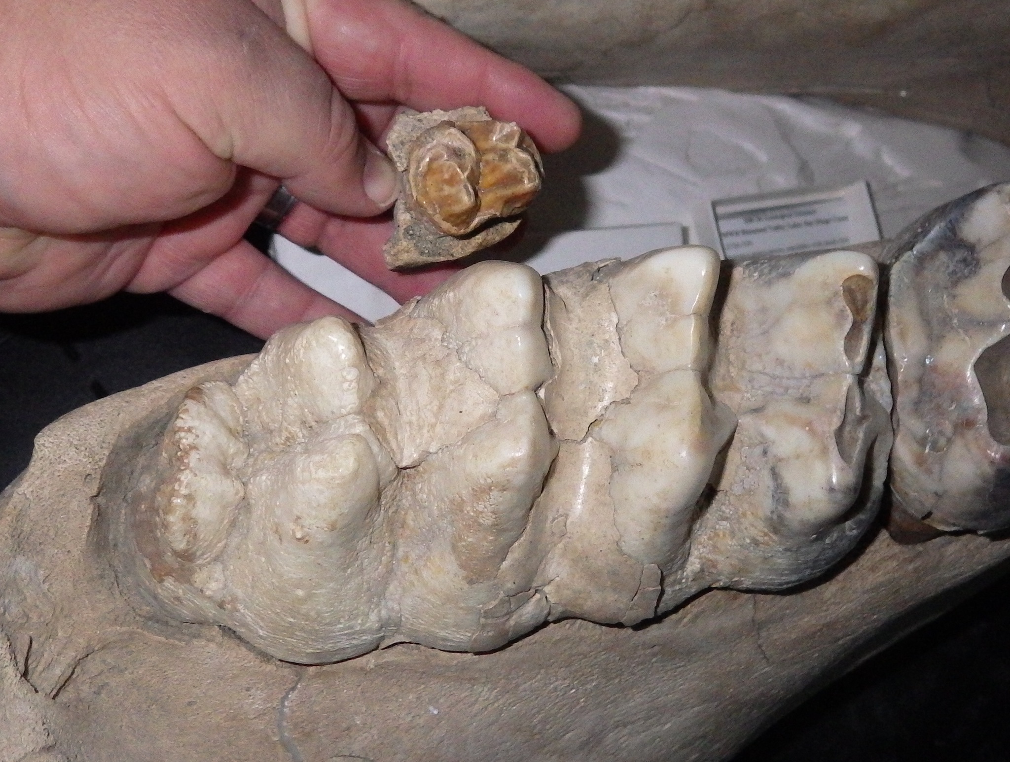 Here's a labial (side) view of the premolar:
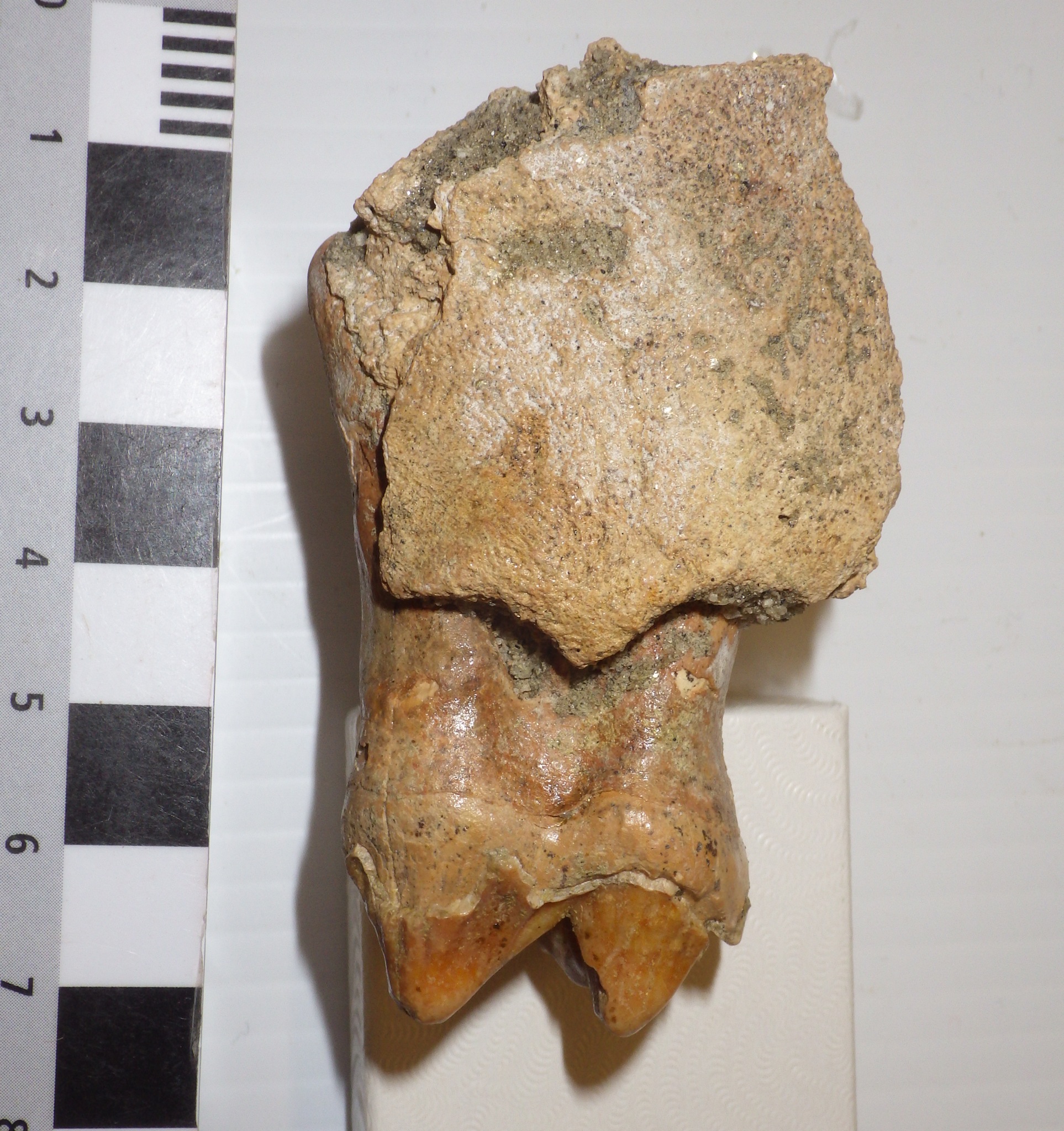 This tooth was discovered near the West Dam in December 1997. It turns out that some other baby mastodon teeth were found at the same site three days earlier. While they have separate numbers in the collection, it's very likely that these teeth all came from one animal. The additional material includes the left maxilla fragment below, shown in labial view with the front toward the left:
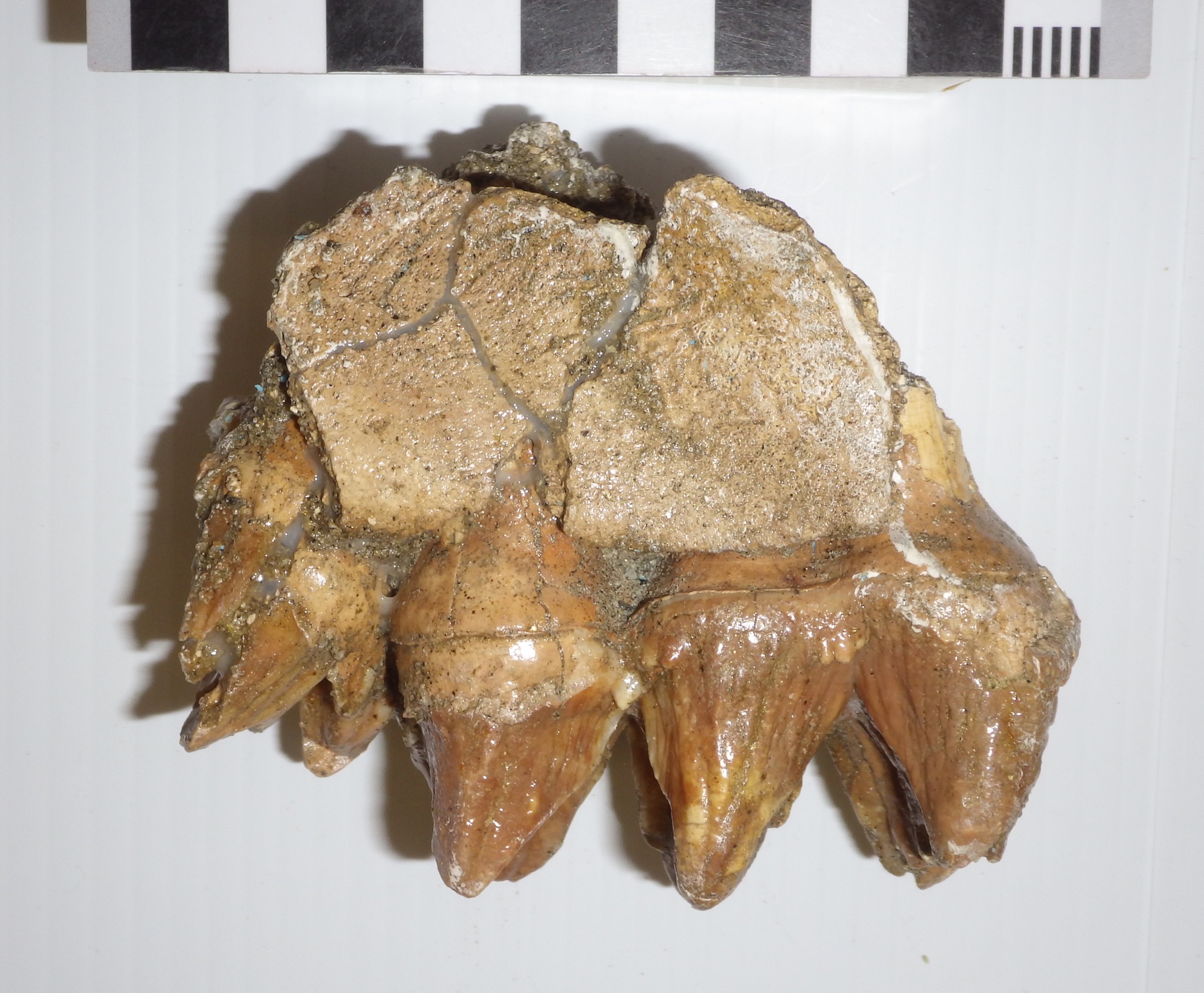 Here's the same fragment in occlusal view, again with the front toward the left:
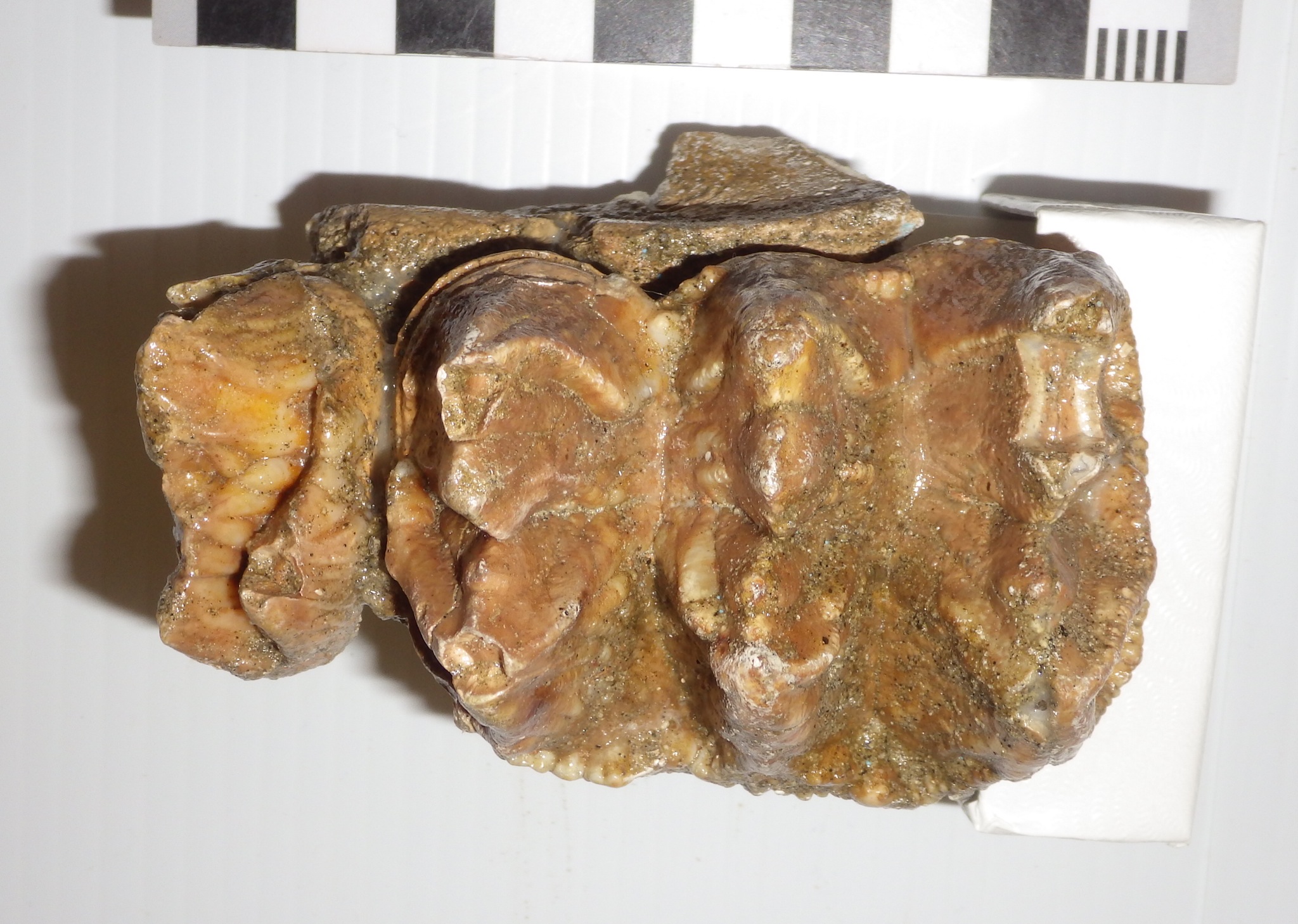 The larger, complete tooth is the fourth premolar (note the three transverse enamel ridges). In front of it is the posterior ridge of the third premolar.The fragmentary right fourth premolar was also collected (occlusal view, anterior to the right):
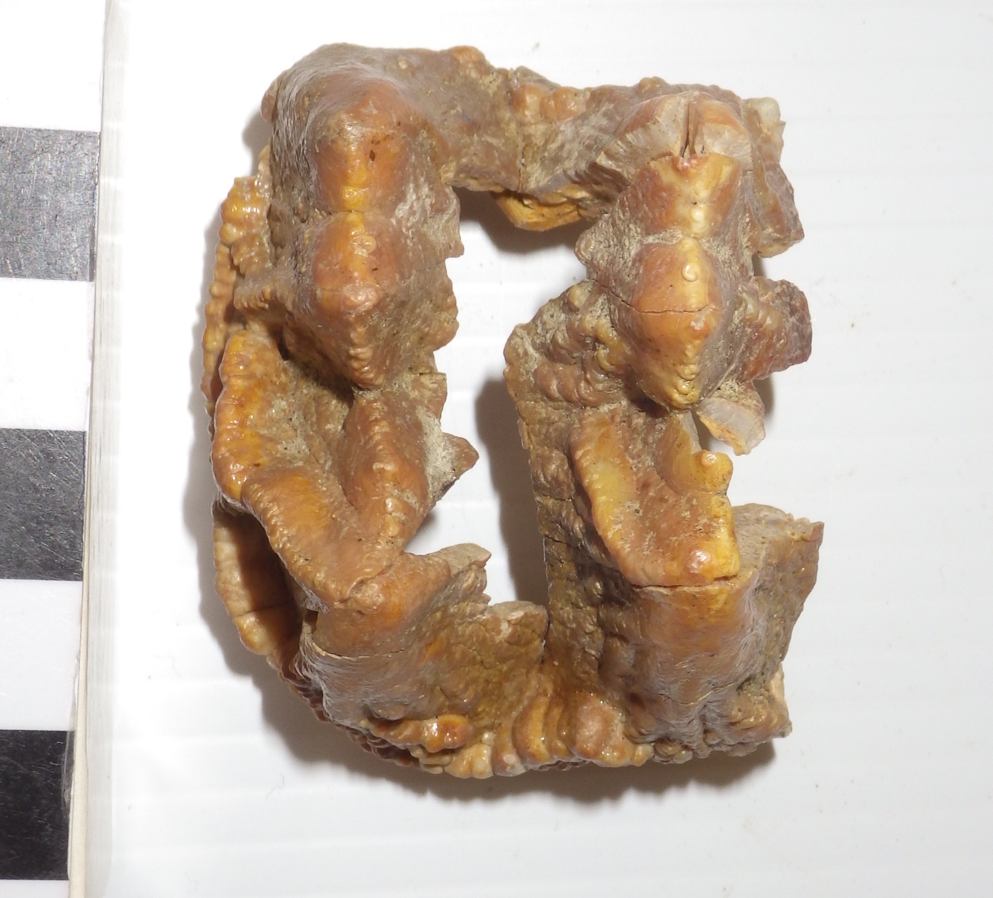 If all these pieces came from one individual (which I think is very likely), we have almost the entire upper left premolar series, lacking only the front half of the third premolar, as well as part of one right premolar.So I called this a baby mastodon, but how young was it? We can estimate the age if we assume that mastodon growth rates and tooth eruption times were similar to those of modern elephants. Mastodonts and elephants are only distantly related, so undoubtedly this introduces some uncertainty, but given their similar sizes and tooth replacement patterns it should produce a reasonable estimate.A key feature to look at is the amount of wear on each tooth. The fourth premolars and the preserved part of the third premolar show no wear at all. But in the second premolar, while the enamel ridges are mostly intact, there is a little wear at the tips (this is most clearly visible in the occlusal view at the top). In African elephants, the second premolar begins to wear when the elephant is about 2 months old, and by the age of 2 years the tooth has worn down and fallen out. Given the light wear present on this tooth, this mastodon was likely no more than 4 to 6 months old!Reference: Green, J. L. And R. C. Hulbert, Jr. 2005. The deciduous premolars of Mammut americanum (Mammalia, Proboscidea). Journal of Vertebrate Paleontology 25:702-715.

