

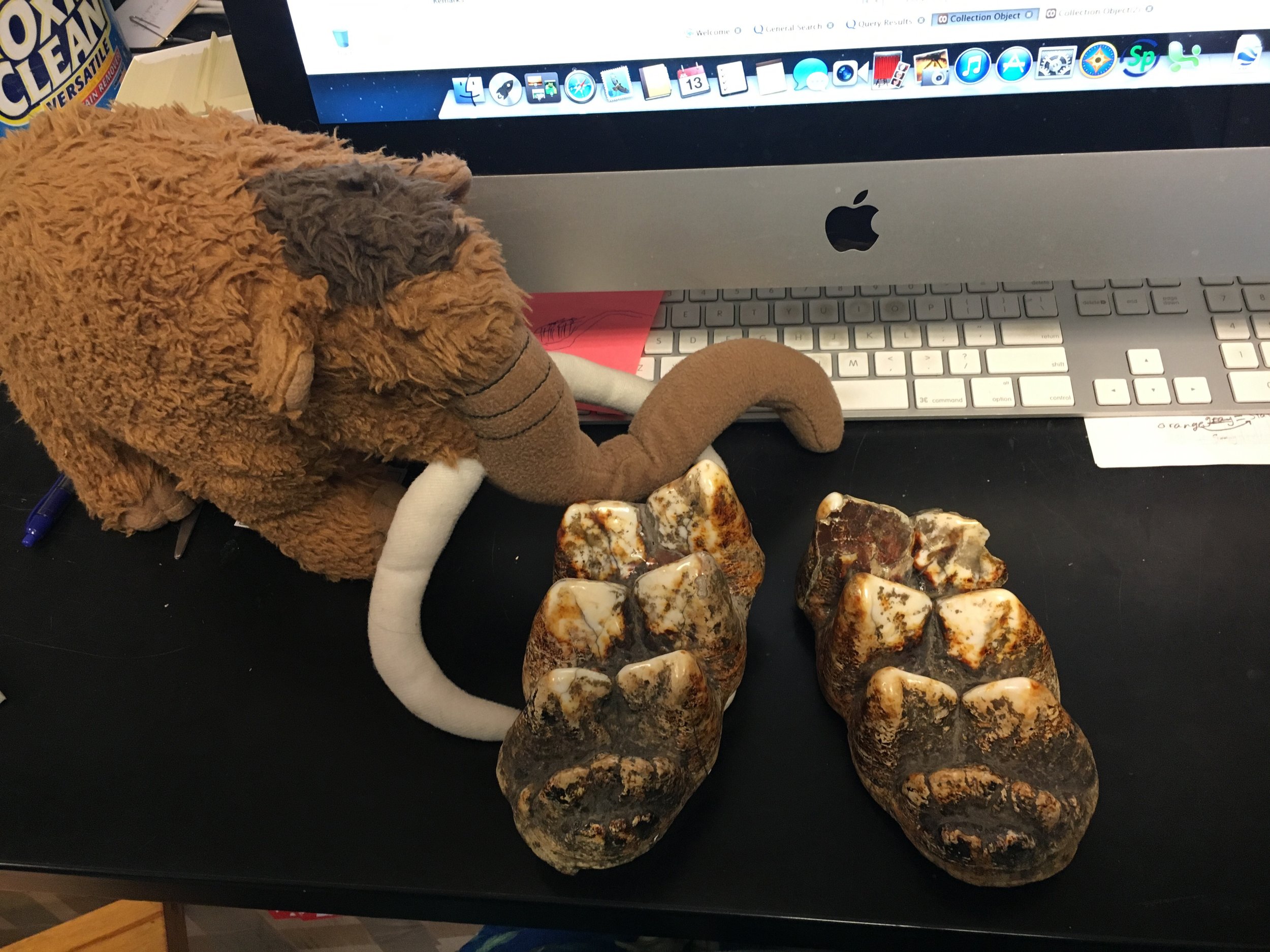
 Yesterday we made our fifth stop on the Mastodons of Unusual Size tour, the University of Wyoming Geological Museum in Laramie. Mastodons get increasingly scarce as you move west, but we had hopes that something would turn up in the excellent fossil mammal collections at the Geological Museum.With the help of Collections Manager Laura Vietti, we tracked down the only two teeth in the Geological Museum's collection, shown above with Max. Both of these teeth are lower third molars, but unfortunately the first lophid is missing on each tooth, so we weren't able to get measurements on either one. Even more surprising, it appears that both teeth are originally from Illinois. With these teeth ruled out, it appears that there are no known mastodon specimens from Wyoming!This is actually interesting news for us, because lots of Pleistocene animals from other species have been found in Wyoming over the years. If mastodons were common there, they should have been found by now, so it seems they were almost certainly absent or at least very rare in Wyoming. Interestingly, when I was planning this trip I contacted the South Dakota School of Mines Geological Museum in nearby Rapid City, which also holds lots of fossil mammals, only to be told that while their collections included lots of mammoths, they didn't have any mastodons at all. It seems that mastodons may have almost entirely avoided the high plains.Yesterday afternoon we left Laramie and headed to Denver for our next stop. More on that to come!
Mastodons of Unusual Size - Joseph Moore Museum
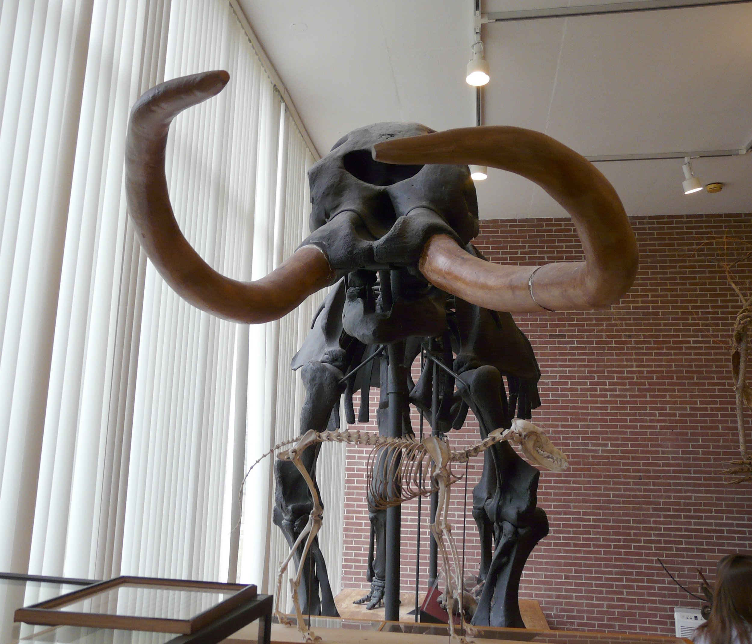 This morning Brett, Max, and I visited the next museum on the "Mastodons of Unusual Size" tour, the Joseph Moore Museum at Earlham College in Richmond, Indiana.The Joseph Moore Museum has a small but significant collection of Pleistocene fossils, with a mounted mastodon composite skeleton (above) as one of the centerpiece exhibits. Unfortunately, the teeth on this specimen are damaged and couldn't be measured, but there were several other mastodon teeth in the collection.
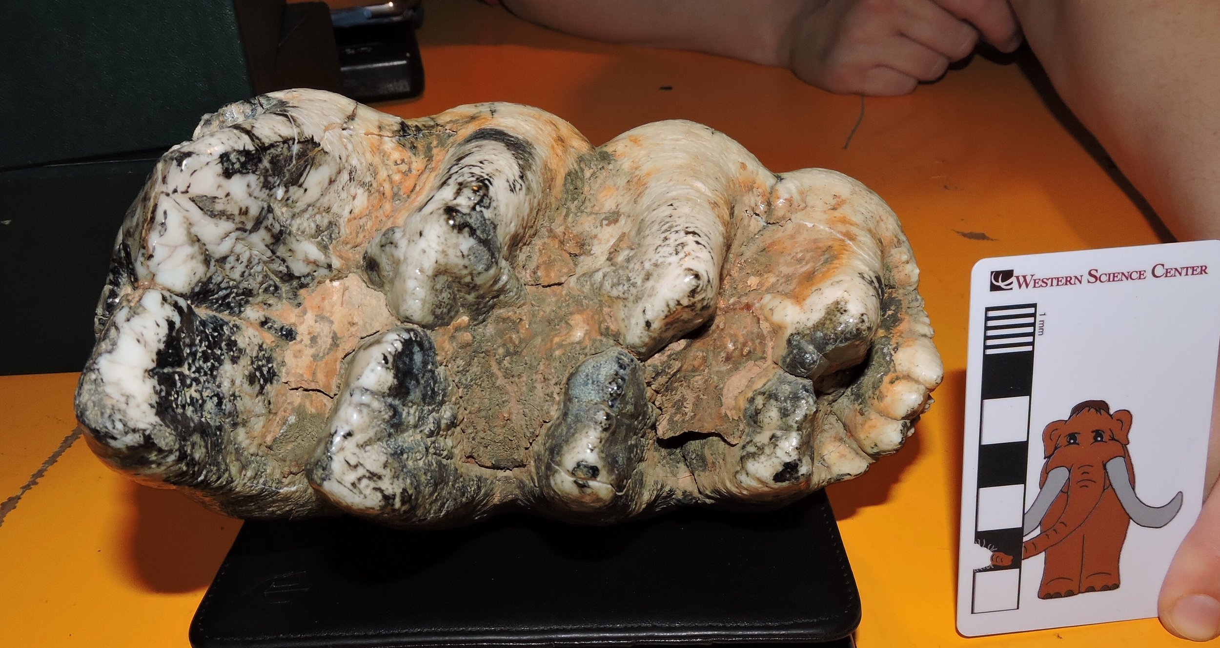 The tooth shown above, from Indiana, is a fairly typical lower third molar, with four lophids (the transverse enamel ridges) and a large posterior cingulid (the bumpy ridge at the back of the tooth that looks like a small lophid on the right in the image). Compare this to from another one in the Joseph Moore Museum collection below, from Ohio:
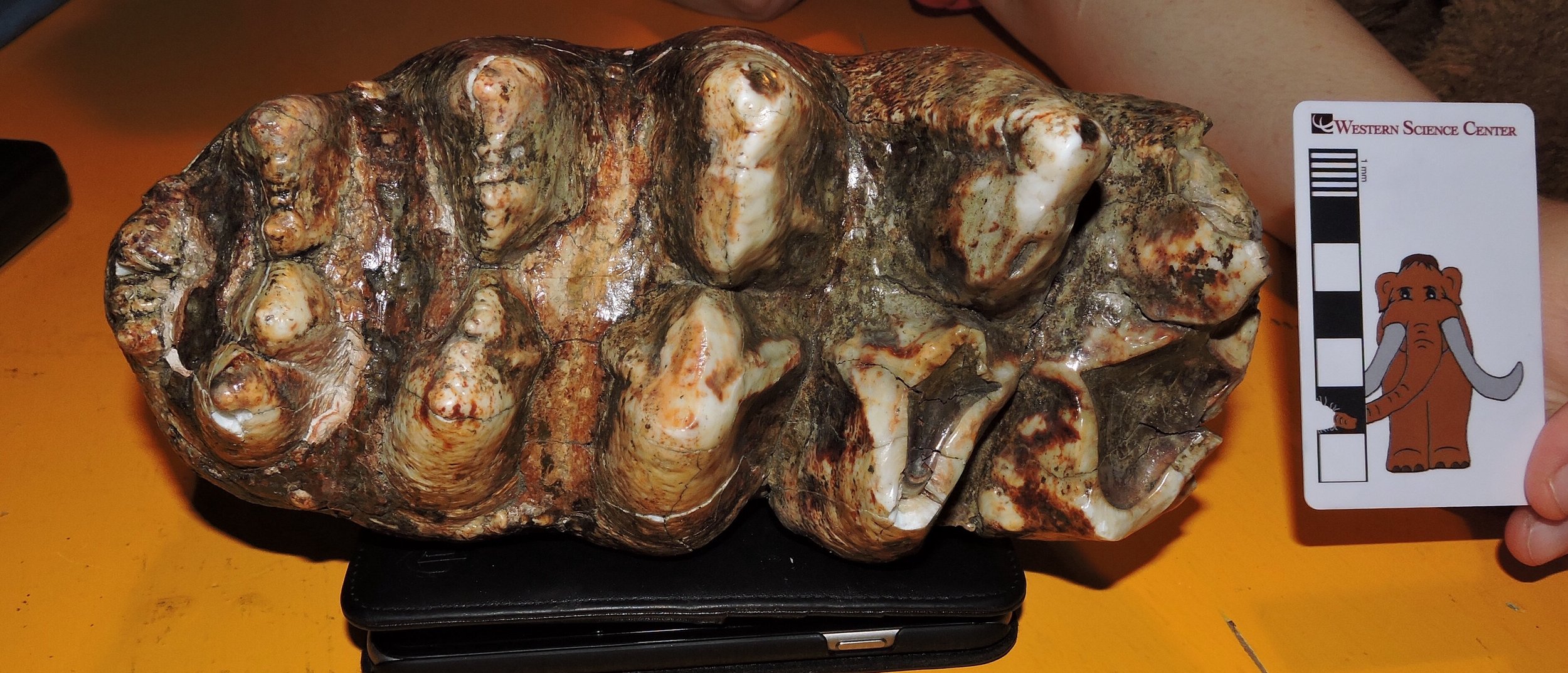 This one is also a lower third molar, but this one has five lophids, and still has a posterior cingulid (posterior is on the left in this image). The five lophid morphology seems to show up in a small percentage of mastodons; we've seen it in only a few specimens so far on this trip, so it nice to find this one at Earlham.We were able to add five teeth to our dataset on this visit. Thanks to JMM Director Heather Lerner for providing access to Earlham's collection.
Mastodons of Unusual Size - Boonshoft Museum of Discovery
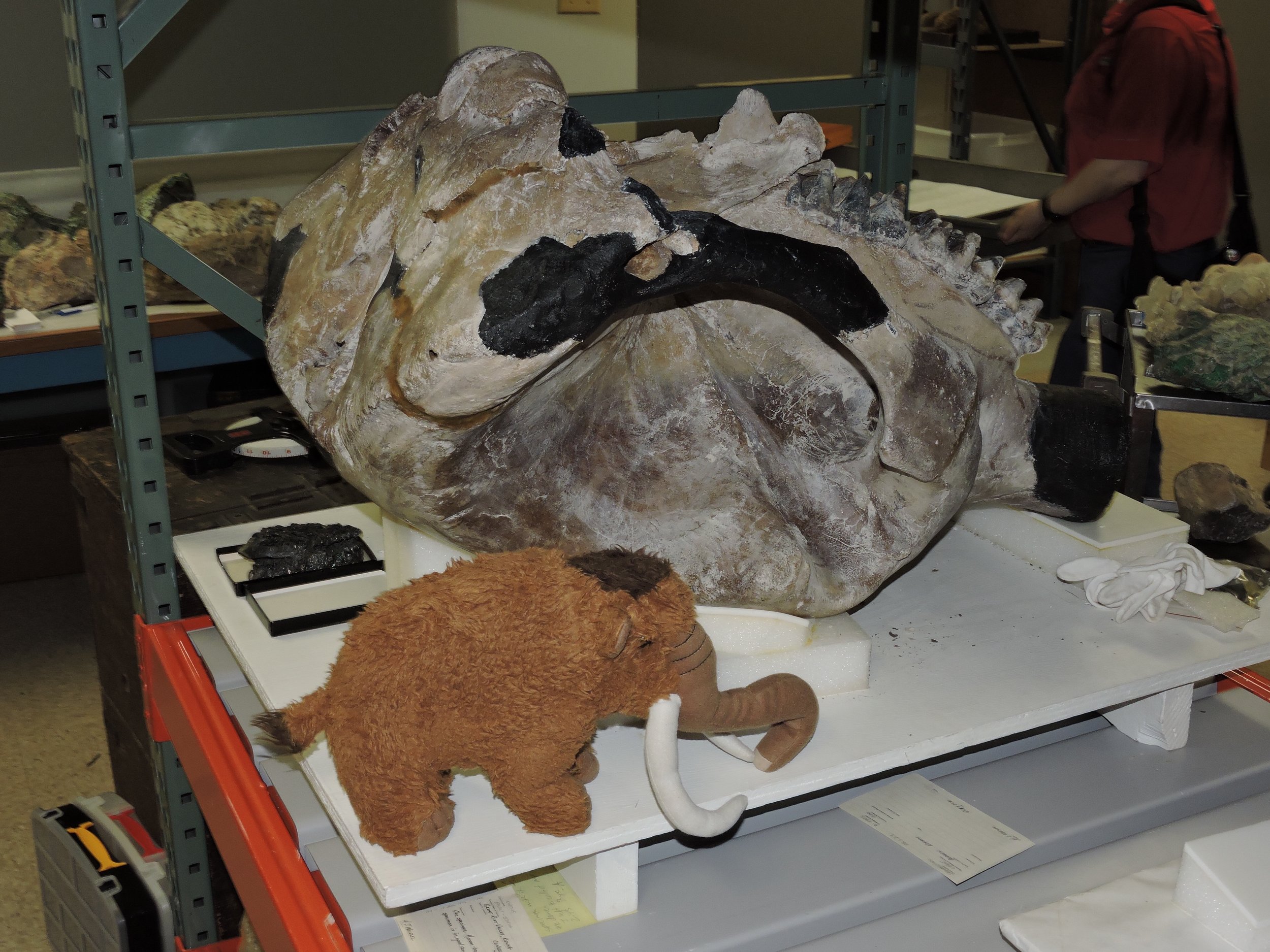 Brett, Max, and I headed back into Ohio this morning for our next stop on the "Mastodons of Unusual Size" tour; the Boonshoft Museum of Discovery in Dayton.The most impressive Boonshoft mastodon is a more-or-less complete skull, shown above with Max. This was a young adult animal, perhaps 16-20 years old based on tooth wear. The first and second molars were erupted and functional, but the third molars had not yet erupted (the tooth eruption stage is very close to that of Xena, the Columbian mammoth on exhibit at WSC). This specimen also includes some postcranial bones, including both femora (below is the right one in anterior view):
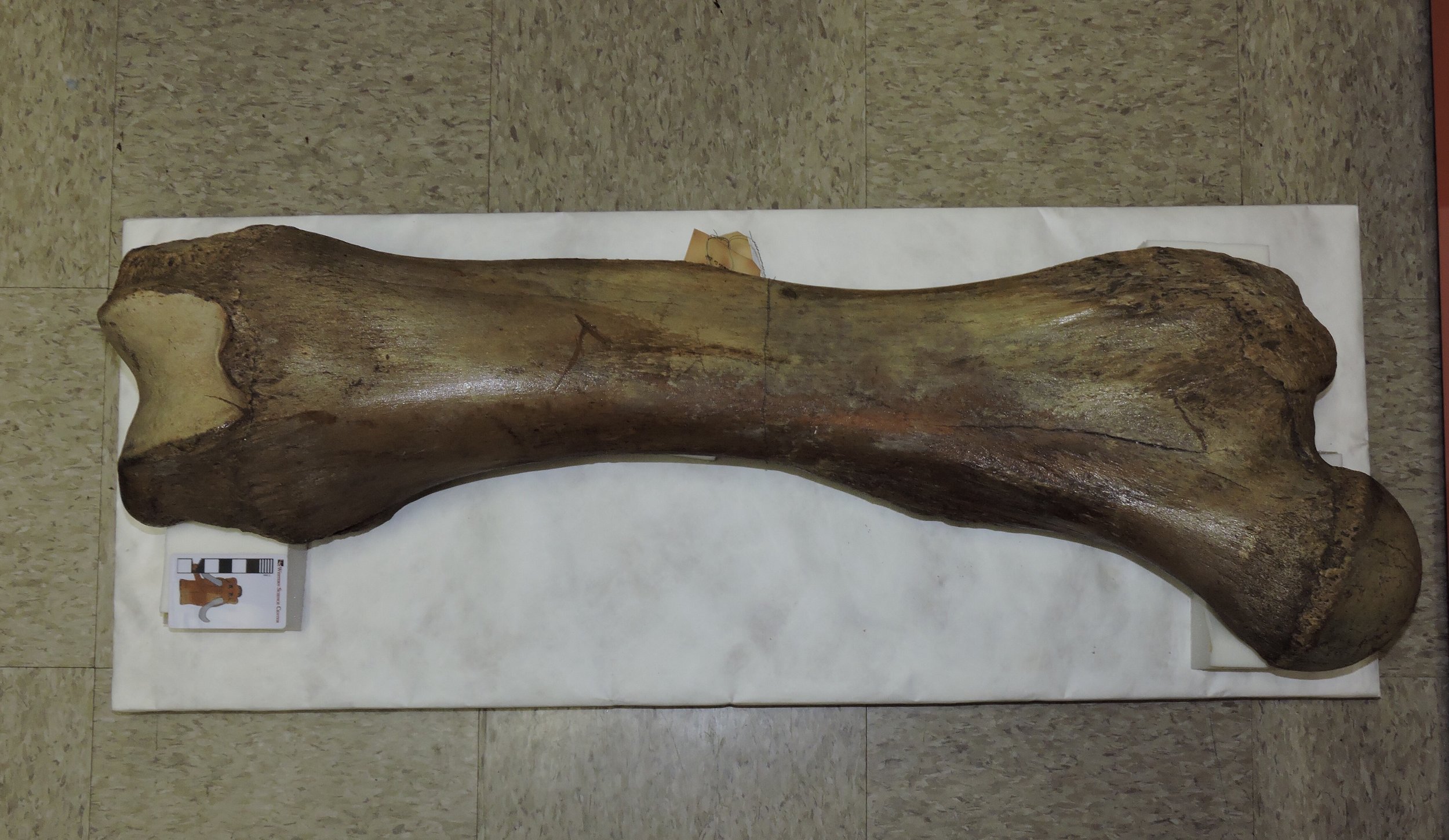 The presence of the femora is nice because they give us a data point for comparing tooth size to body size.Below is simultaneously the most intriguing and frustrating specimen we saw today:
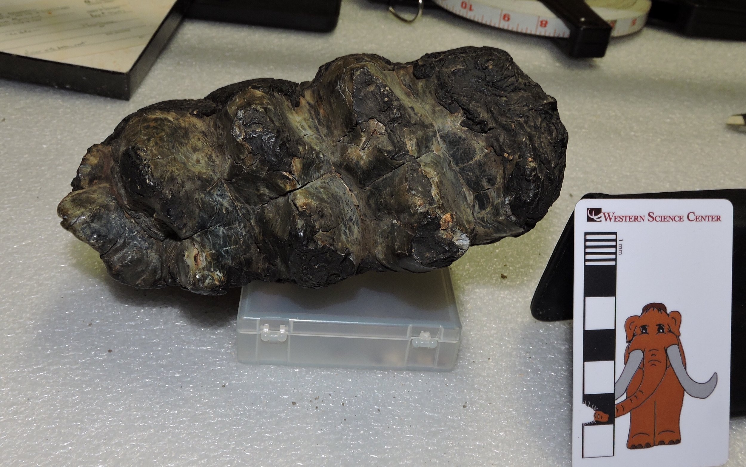 This small lower third molar has both the size and proportions we would normally expect to see in a California mastodon. So, is this an Ohio specimen that looks like a Californian one? Maybe not! The Boonshoft has a few specimens acquired nearly 100 years ago from Rancho La Brea. This tooth has black residue on it that looks a lot like Rancho La Brea sediment. But...the specimen has no locality information, so we can't confirm where it's from (at least not without some expensive chemical testing)!We examined 17 teeth in the Boonshoft collection, which came from nine individuals; 4 of these teeth didn't have locality data and probably can't be included in our study. Most of the specimens are from Ohio, but we also examined a lower jaw from Florida. The addition of a skull with associated femora is significant, and prior to our Cincinnati and Boonshoft visits we had almost no Ohio or Kentucky specimens in our database. Thanks to Boonshoft's Anthropology Curator Bill Kennedy (below, with Max) for helping us with access to the collections.Tomorrow "Mastodons of Unusual Size" continues with our next stop.
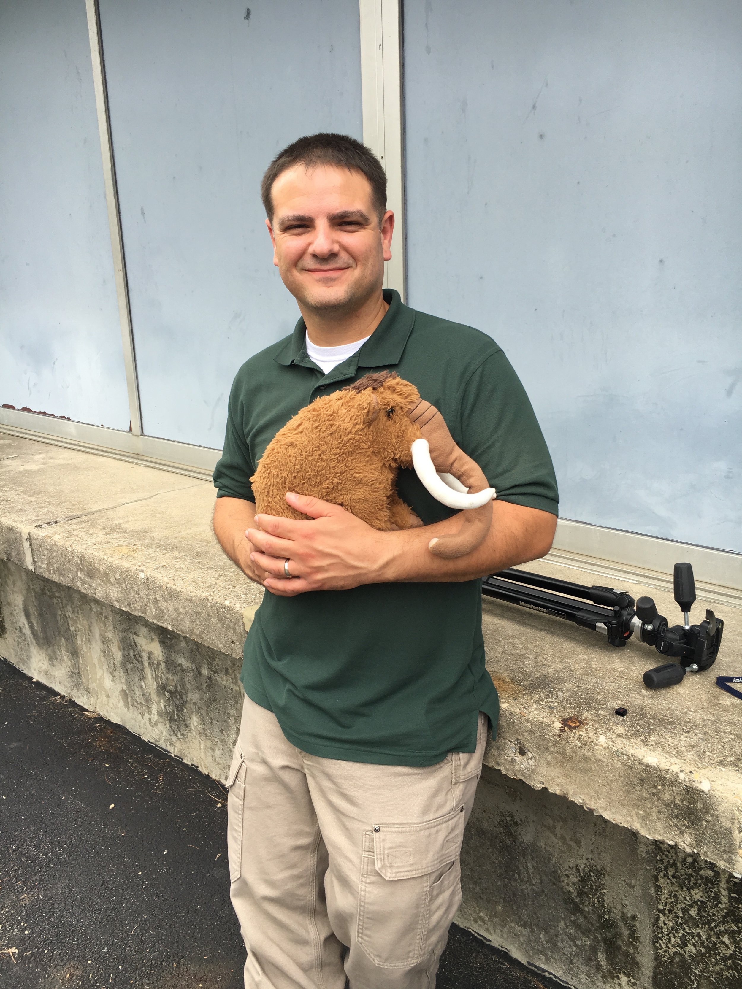
Fossil Friday - mastodon thoracic vertebra
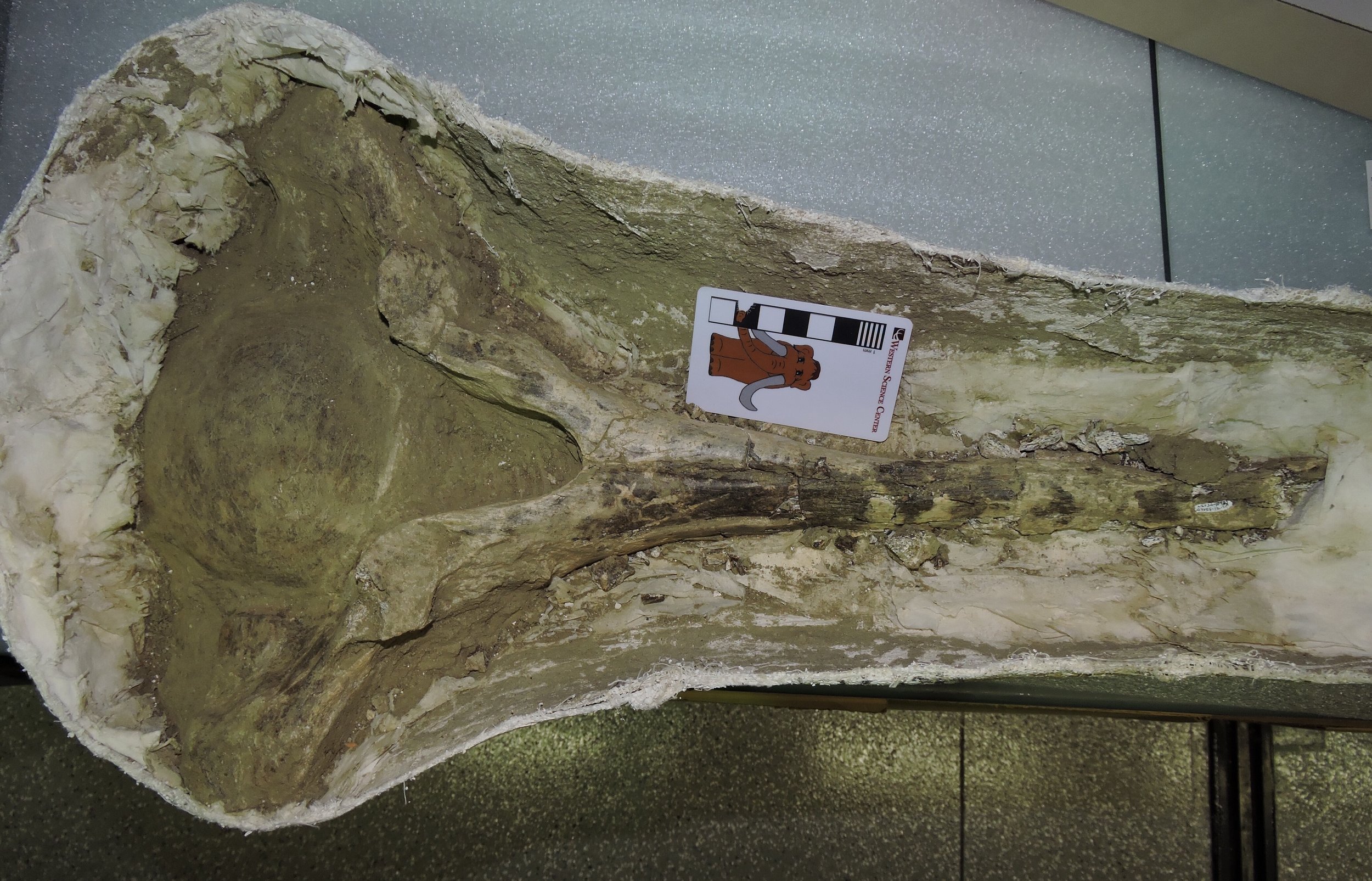 As I'm in the middle of our Mastodons of Unusual Size data-collecting trip, my Fossil Friday post will have to be a short one. But, in keeping with our road trip topic, it will of course feature a mastodon!The specimen shown above is an anterior thoracic vertebra, shown in anterior view. The image is turned sideways to fit better on the screen, so the bottom of the vertebra is on the left. The bone is still in its field jacket, and has only been partially prepared.Thoracic vertebra in mammals are the ones that articulate with the ribs, and the articulations are visible on the ends of the processes projecting from each side of the vertebra. The tall neural spine extends from the top of the vertebra (right in the photo). The neural spines serve in part as muscle attachment points for muscles that hold up the head and help support the front legs. In quadrupedal animals with large, heavy heads, such as mastodons, the anterior neural spines are very long, providing more surface area and better leverage for those muscles.This vertebra was collected from the Diamond Valley Lake East Dam, close to where the Western Science Center is now located.
Mastodons of Unusual Size - Cincinnati Museum of Natural History
 After leaving Baton Rouge, Brett and I (accompanied by Max the Mastodon) headed north to our second data collection stop: the Cincinnati Museum of Natural History. The Overmyer mastodon from Indiana is one of the premier CMNH mastodons (Max is shown above with the lower jaw). This specimen has actually been published, so much of the data we needed was already available, but we were able to get measurements on postcranial bones and photos of the lower teeth.All told, we took measurements on 2 femora and 19 teeth, although 6 of the teeth have limited locality information. Several of the teeth were quite large, although none were as big as the monster tooth from Baton Rouge we saw on Tuesday. The lower jaw below was particularly interesting:
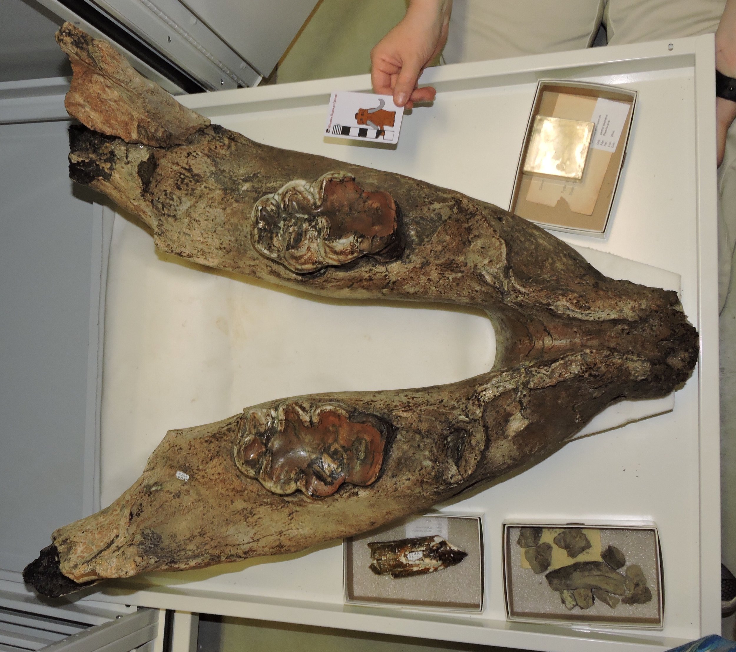 This specimen only has third molars, which are heavily worn across their entire length; the second molars have fallen out and the sockets have grown over. This was a really old animal when it died, very likely past 60 years old.The lower third molar below, from Kentucky, is also noteworthy for a couple of reasons:
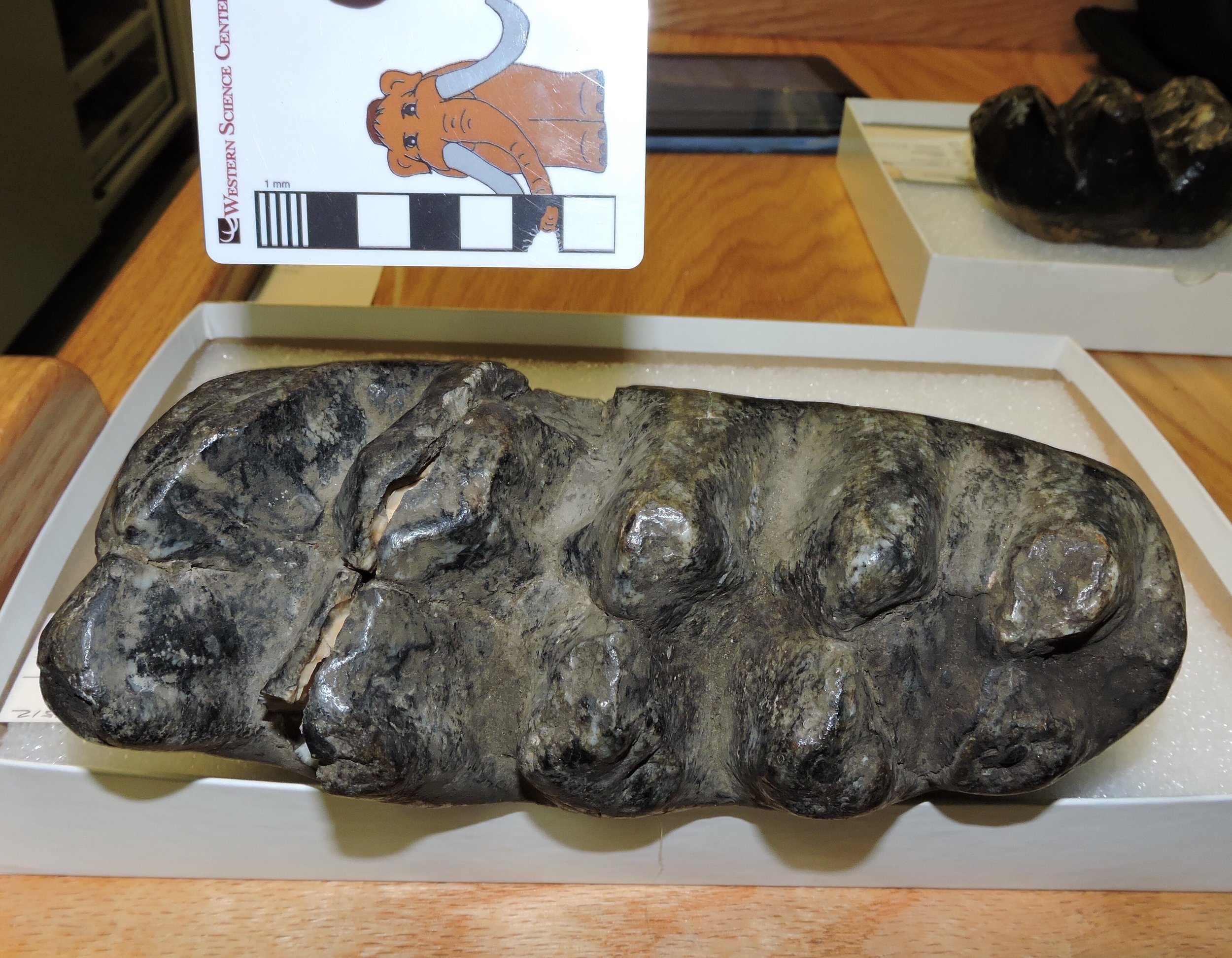 First, it has five distinct lophs on the crown, instead of the four we usually see in Mammut third molars. Second, this is a remarkably narrow tooth; it's actually even narrow by California standards. So far we've only seen a handful of teeth with these proportions outside of California. That said, this is still a large tooth, much longer than any California tooth.After leaving Cincinnati we headed north to Richmond, Indiana, where we'll spend the next few days while visiting several area museums.
Mastodons of Unusual Size - LSU
 Today Brett and I made our first stop on our experiment.com - funded trip to collect data on mastodon tooth proportions: Louisiana State University.I received my Ph.D. from LSU in 1998, and this was only the second time I've been on campus since graduating. My graduate advisor, Dr. Judith Schiebout, is still at LSU, and Dr. Ting Suyin, who was a graduate student the same time I was there, is now LSU's vertebrate fossil collections manager:
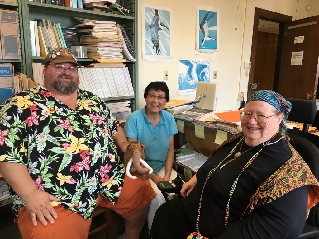 Of course, our real reason for being here was to measure mastodons. We collected measurements and photographs for 17 teeth, although only 13 are suitable for the database (the others are too poorly preserved for a complete set of measurements).While the tooth proportions of the Louisiana specimens were about what we would expect for teeth from the Gulf Coast (relatively much wider than California specimens), I was struck by how large some of these teeth are. The tooth at the top, an upper 3rd molar from the Tunica Hills region, is a pretty large tooth that is both longer and wider than any California tooth. But even it was dwarfed by this monster from Baton Rouge:
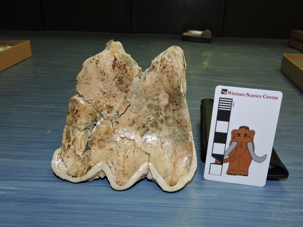
 These are occlusal views of the upper right 2nd molar and lower right 3rd molar; the 3rd molar is damaged, so we don't have the full width. These teeth are absolutely enormous. The 3rd molar is the second longest tooth in our database, and is more than 2.5 cm longer than any California m3. The M2 is 117.7 mm wide, the only M2 in our data set that's more than 100 mm wide!Below, Brett is holding a maxilla fragment with an M3 from another Louisiana specimen we measured today:
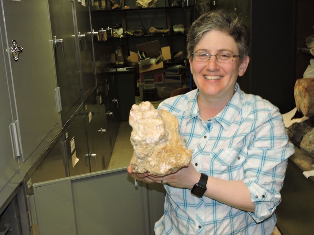 This specimen is special to me, because Brett and I led the excavation that recovered it. Brett and I were married in 1994, and after our honeymoon we drove to Baton Rouge where I was enrolled as a graduate student and Brett was starting a job as a lab technician for Dr. Schiebout. Immediately after we arrived we were called out to Angola, Louisiana to examine possible fossils that had been uncovered during a construction project at the Louisiana State Penitentiary. The remains turned out to be this specimen, and over the next several weeks Brett and I led an LSU crew on an excavation at the prison (this remains the only mastodon I've ever personally excavated, and the only time I've run an excavation inside a prison!). At the time we received a good deal of local media coverage, which only intensified when during the course of the excavation we discovered an unmarked and eroding 1800's-era cemetery directly above the excavation site. The photos below originally ran in the Baton Rouge Advocate, showing archaeologists working on the cemetery site at the top of the hill while we paleontologists simultaneously worked on the mastodon below them, and a closeup of the archaeological site:
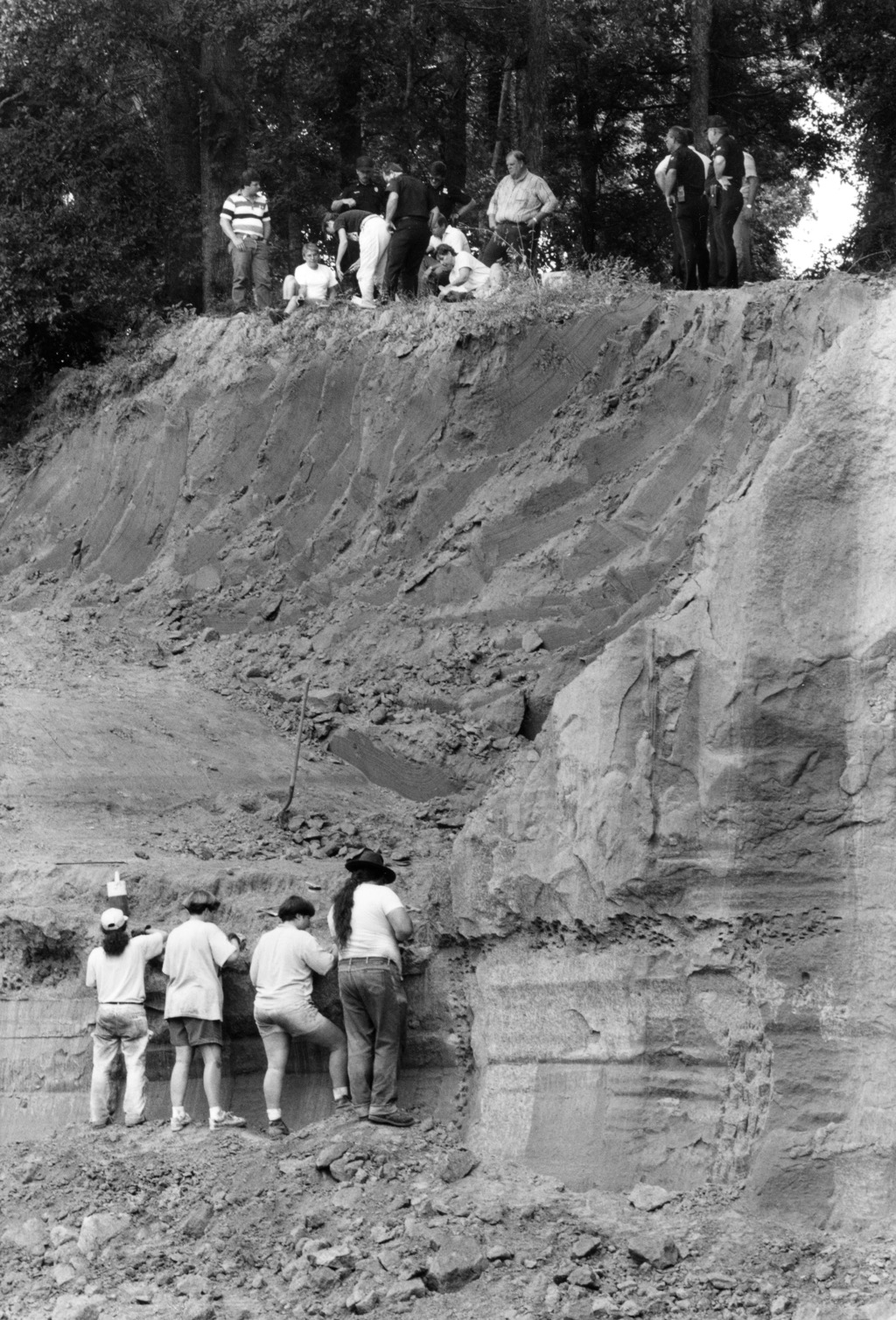
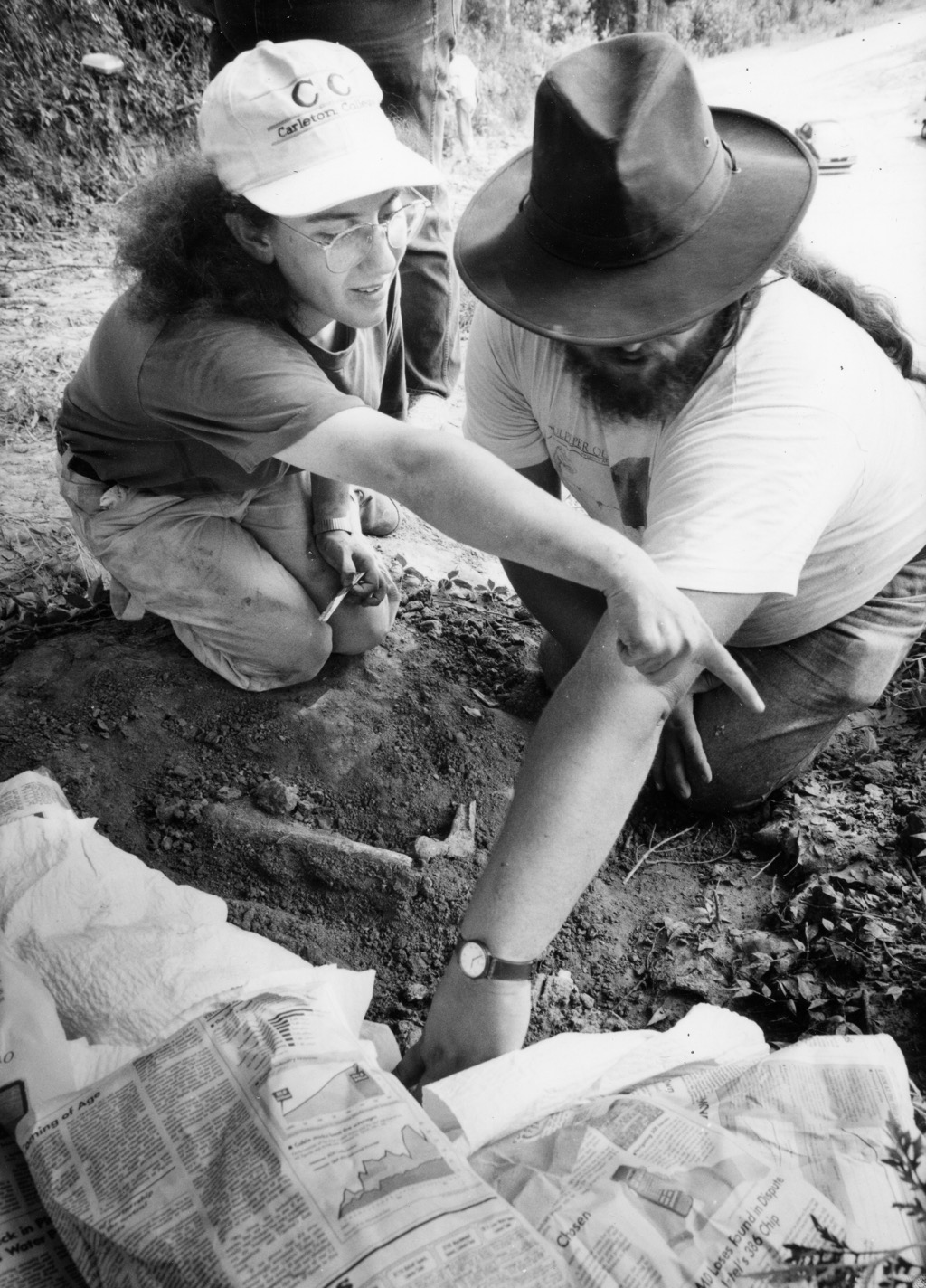 (That really is Brett and me, 22 years ago!)After documentation, the human remains were reburied in a new location. The mastodon remained in jackets until after I graduated, and was finally prepared sometime after 2000. Today was the first time Brett and I had seen the specimen since excavating it in 1994!Tomorrow we leave Baton Rouge and head north to visit more museums. Follow along here and on Max's Twitter feed (@MaxMastodon) and at #MastodonsOfUnusualSize.
Capitan Reef Complex
 As Brett and I headed east over the weekend on our way to collect mastodon data, we made a few detours to examine geologic features and collect fossils for the museum.At a roadcut just east of El Paso, Texas (above) we spent 30 minutes examining and collecting small fossils:
 Notice the oblong dark splotches above the scale bar, that are just a few millimeters long. Here's a closeup of one of them:
 These fossils may be tiny, but they're actually giants within their group. These are shells (or "tests") from fusilinids, a type of foraminifer. Foraminifera are protists, so these are shells from single-celled organisms!Fusilinid foraminifera first show up in the fossil record during the Silurian Period, but they reached their heyday during the Permian Period, when they were present in vast numbers in shallow marine waters; the rocks in this area are Permian rocks. The fusilinid party wouldn't last, however. After surviving more than 150 million years, the entire fusilinid line went extinct at the end of the Permian Period during the huge Permo-Triassic mass extinction when 90% of the species on Earth were wiped out.Continuing east, this imposing structure rose up out of the desert in front of us:
 This is the southern end of the Guadalupe Mountains, with a huge limestone cliff called "El Capitan" forming the summit:
 (See a Gigapan we shot of El Capitan here.)El Capitan is part of Guadalupe Mountains National Park, so accompanied by Max we made a brief stop at the visitors center:
 El Capitan is the namesake of a feature known as the Capitan Reef Complex, a huge Permian barrier reef which is formed in part by the Guadalupe Mountains (Carlsbad Caverns in New Mexico is also part of the reef), and which surrounds a depression called the Delaware Basin. This reef system was made up largely of algal mounds called stromatolites and oncolites, such as the ones below (all the closeup images below are specimens on display at the park visitor center):
 Sponges were also major contributors to the reef system:
 As with most reefs, there was a vast array of other organisms living on the reef, including brachiopods:
 ...crinoids:
 ...bryozoans:
 ...and bivalves:
 ...as well as rugose corals, fusilinids, cephalopods, fish, and other animals.So how did this reef end up in Texas? The origin of the reef is tied in tightly with plate tectonics. In the period immediately before the Permian, the Pennsylvanian Period, a major continental collision began to take place between Gondwana (a continent made up in part of what is today South America, Africa, Australia, Antarctica, and India) and Laurasia (which today forms North America, Europe, and most of Asia). At the end of the Pennsylvanian Period and the beginning of the Permian Period, the Gondwana-Laurasia collision formed the Ouachita and Appalachian Mountains. A large mountain range such as the Ouachitas is so heavy that it pushes down the surrounding continent, forming a low area called a foreland basin. This basin can be hundred or even thousands of meters deep, and if it connects to the ocean it will fill with sea water. The Persian Gulf is a modern example of such a basin. Below is a map from Ron Blakey showing the North American part of Laurasia during the middle Permian (link to the original map):
 And below is a version I've marked up with the locations of the collision, the foreland basin, and the Capitan Reef Complex:
 Outside the reef are extensive shallow marine sediments, often with fusilinids and other marine fossils, such as the ones we examined near El Paso. Inside the reef, water flow is restricted and evaporation rates are high. This results in the deposition of salt, gypsum, and other minerals that dissolve in seawater, leaving evaporite deposits:
 Since foreland basins always have a mountain range nearby (which is what causes them to form in the first place), they rapidly fill with sediment eroded from the mountains and with limestone produced by marine organisms and evaporites precipitated from seawater. By the end of the Permian Period the Delaware Basin had filled with sediment more or less to the top of the Capitan Reef, and the reef was exposed as dry land. It partially eroded and the remnants were buried, but about 200 million years later another tectonic event, the Laramie Orogeny, formed faults that brought part of the reef, including the Guadalupe Mountains, back to the surface. Subsequent erosion removed many of the softer evaporites and formed caves such as Carlsbad Caverns.Reference:Much of the information in this post was based on this report (pdf):Standen, A., S. Finch, R. Williams, B. Lee-Brand, and P. Kirby. 2009. Kapitän Reef Complex Structure and Stratigraphy. Texas Water Development Board Contract Number 0804830794, 53 pages plus appendices.
Fossil Friday - mastodon molar
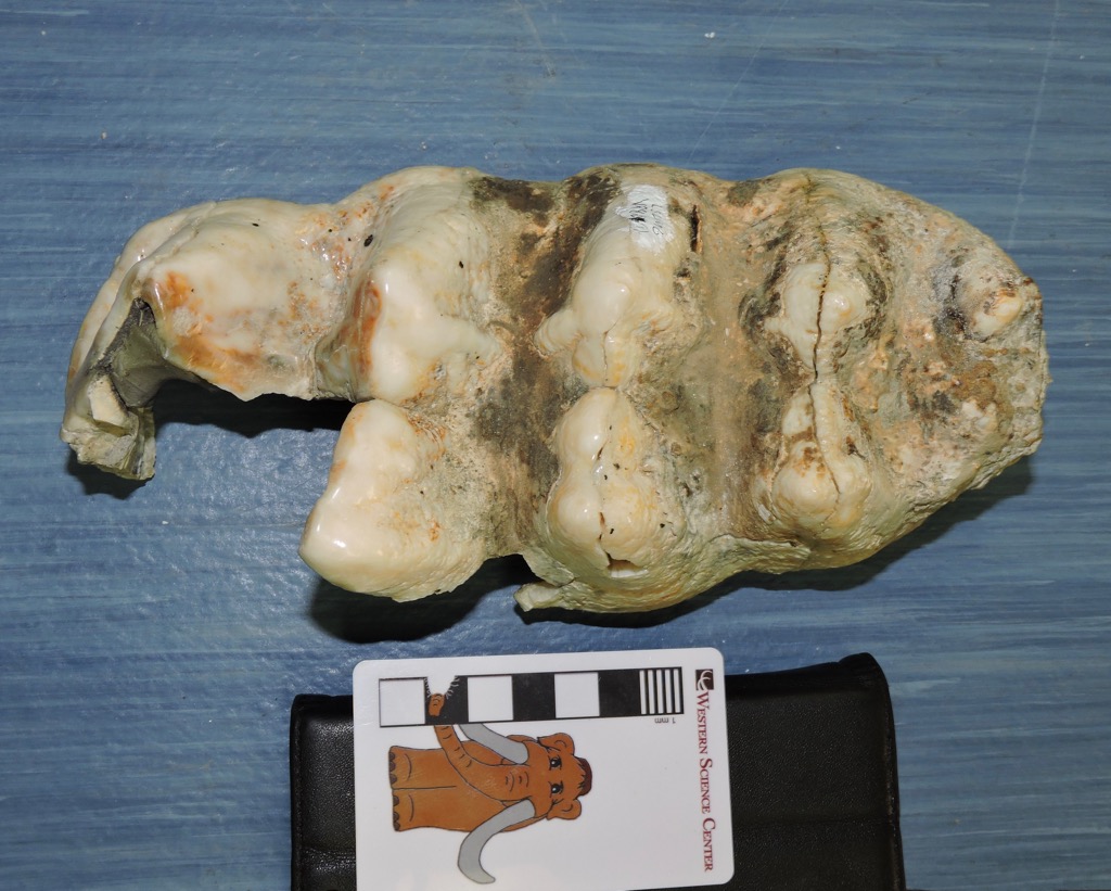
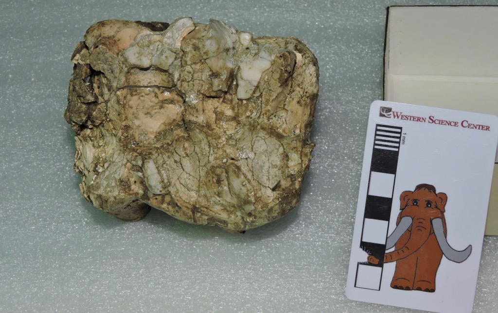 Sometimes the fossils we recover are a little worse for wear. The remains may have deteriorated before burial, been damaged or deformed by sediment weight, structural forces, or groundwater after burial, weather out of the sediment and get eroded before discovery, or damaged during the act of discovery or excavation (I've collected numerous fossils that were "discovered" by bulldozers!). Sometimes it seems amazing that any fossils reach us at all. And yet, even these damaged specimens can be significant.The rather amorphous lump above is actually a mastodon tooth from Diamond Valley Lake, seen in occlusal view (the chewing surface). If you don't look at mastodon teeth on a regular basis, it might be a little difficult to recognize at first, because a lot of the enamel is missing. It's much more recognizable in lateral view, although the damage to the crown is still obvious:
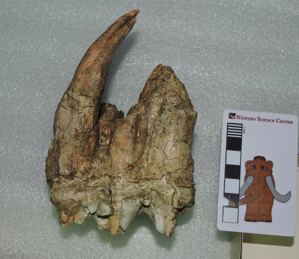 Even with the imperfect preservation, there's quite a lot we can say about this tooth. The lophs (the enamel ridges on the crown) are nearly perpendicular to the long axis of the tooth, suggesting that this is an upper rather than a lower tooth. The curvature of the roots indicates that the anterior side of the tooth is on the left in the photo above. Upper mastodon teeth wear more rapidly along the inside edge (the lingual side) than along the outside edge (the labial side), so that makes this an upper left tooth. There are three lophs present so that limits the tooth to either the fourth premolar, the first molar, or the second molar (the second and third premolars only have two lophs each, while the third molar has four or sometimes five lophs). From P4 to M2 the teeth get progressively larger, and this tooth is pretty large. Combining all these observations, the tooth is the upper left second molar.But wait, there's more! Even though a lot of the enamel is missing, there's enough to tell that the tooth was fairly heavily worn, so it had erupted before this animal died. But the crown wasn't completely worn away; there are still grooves separating the lophs, at least on the labial side, so this tooth was probably still in use when the animal died. For example, Max's 2nd molars, which are still in his jaws, are more worn than this one. That means the mastodon was a young adult when it died. This is supported by the long, intact root, indicating that the tooth had not been shed. Instead, this tooth was still in the mastodon's skull when the animal died, and the skull was broken up at some point, probably before burial.Finally, there is actually enough of this tooth preserved that we will be able to include this specimen in our study of mastodon tooth proportions, since we can get good crown length and width measurements.Brett and I leave this morning on our museum tour to collect measurements on mastodon teeth from other areas, visiting at least five museums across the central U. S. during the next two weeks. I'll be posting updates about our trip on this blog and on experiment.com/mastodons and Twitter (@AltonDooley), as well as on the museum's Instagram and Facebook pages. Max's alter ego is also accompanying us, and will be posting his own updates on his Twitter feed (@MaxMastodon).
Fossil Friday - mastodon mandible
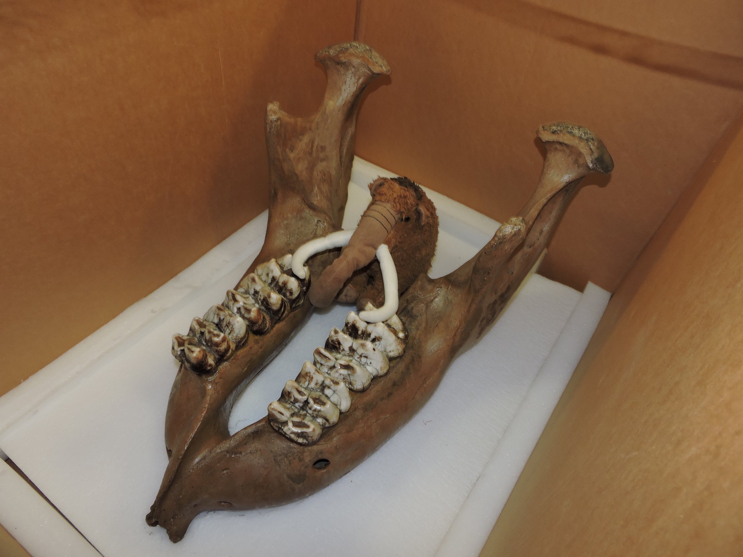
 In a week Brett and I head out on our experiment.com-funded road trip to collect data on mastodon teeth. The major goal of this project is to collect enough data to better understand what's going on with mastodon tooth proportions, especially with respect to the apparently unusual California mastodons.The nicely preserved lower jaw shown above is a WSC specimen recovered from the San Diego Canal near Diamond Valley Lake, and which was included in our recently-closed "Stories from Bones" exhibit. Below is a dorsal view:
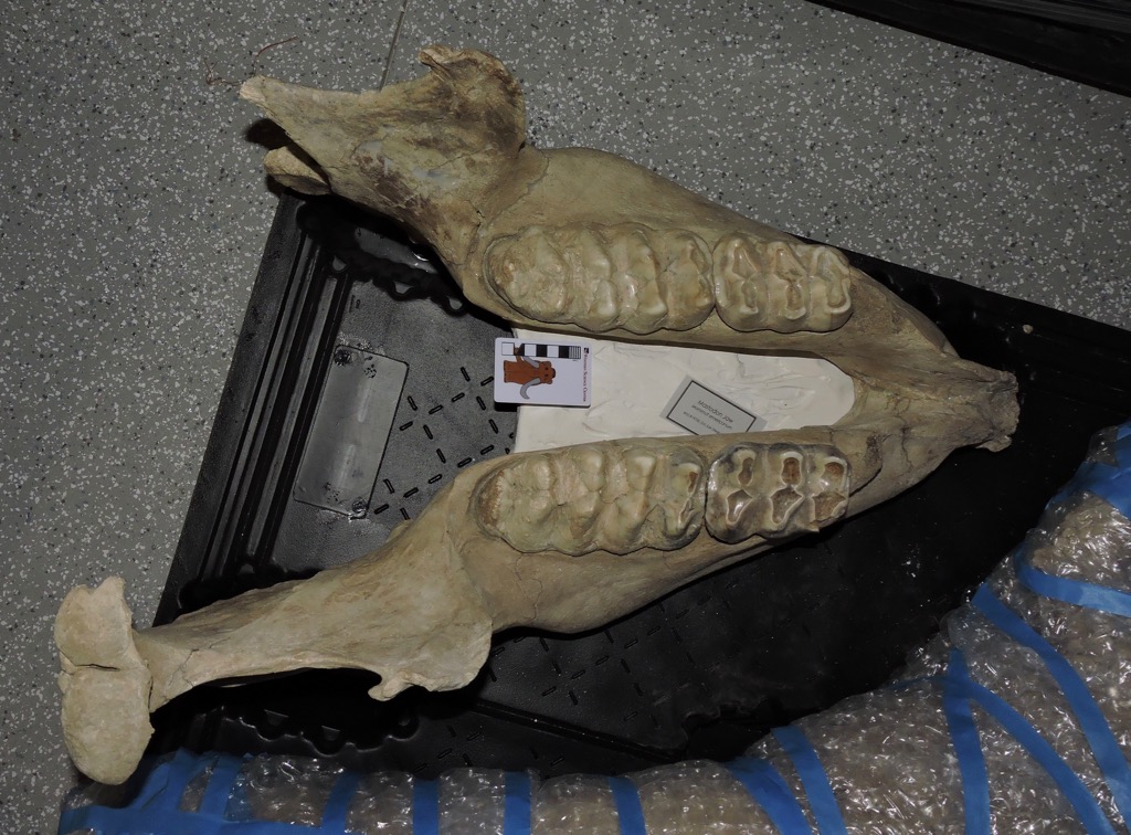 This animal was a little smaller than Max, and based on tooth wear, a bit younger. While the first molars are lost, the third molars are just coming into wear, so this animal was likely in its early 20's.This specimen is of particular interest in our project, because it's an outlier. So far, this mastodon has the widest lower third molars of any California mastodon we've measured, and it's not really even close. The left molar on this specimen is almost 89 millimeters wide, while the next largest California specimen is just over 83 mm (the right molar is a bit narrower, but is still almost 85 mm). This is the only California specimen we've seen that has m3 proportions that approach typical Florida or Indiana specimens.The outlier status of this specimen is even more noticeable because, of the 14 California mastodons with m3 measurements currently in our database, 5 have m3 widths between 82.9 and 83.1 mm. That width seems to be a sharp line that California mastodons almost never cross, with the exception of this one specimen. (As an aside, gigantic Max's m3 is only 76.6 mm wide, one of the narrowest in our data set).As we examine additional California specimens, it's likely we'll eventually find other mastodons with teeth that are comparable to this specimen. But for now it stands out as the only California mastodon that has tooth proportions that approach those of eastern specimens.
Fossil Friday - horned lizard spike
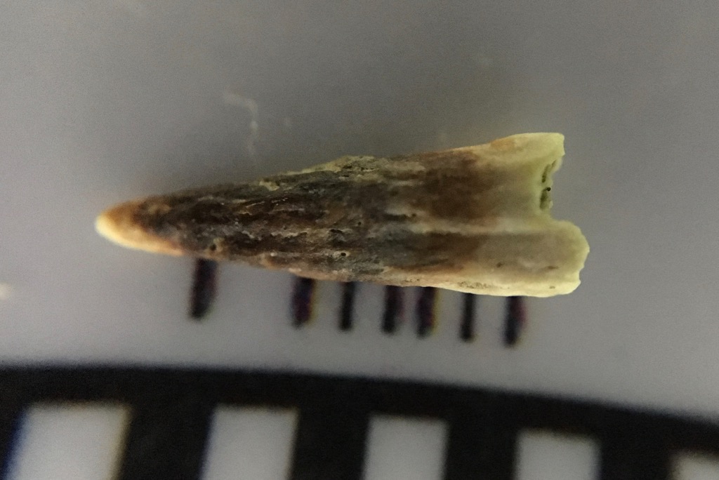 One of the things I find most intriguing about the Pleistocene is how, in spite of the impressive megafauna, it was so similar to the modern world. As I've mentioned in other posts, as with many other Pleistocene sites the fauna at Diamond Valley Lake is in many ways a lot like what we see in southern California today.The tiny fossil shown above (that's a millimeter scale) may look like a tooth at first glance. But the pitted texture is a giveaway that this is a bone, not a tooth. This is a horn from a horned lizard of the genus Phrynosoma, specifically one of the horns that projects from the parietal bone in the skull, visible below in this specimen of Phrynosoma cornutum that Brett photographed in Texas:
 There are a number of different species of horned lizards that are each endemic to different parts of North America, and one of the ways they differ is in the size and shape of their horns. Compare the horns on the Texas lizard above to those on Phyrnosoma douglasii from South Dakota:
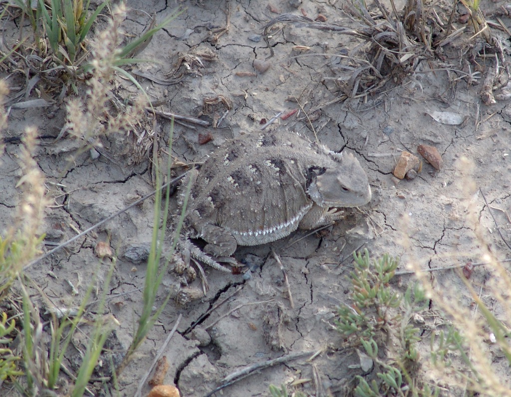 The southern California horned lizard species is Phrynosoma coronatum, which I've never had the opportunity to photograph in the wild. Its skull, however, is somewhat similar to the Texas species with large parietal horns, as can be seen in this comparison image.Horned lizards live in relatively arid areas, which seems to be a common theme with the small Pleistocene animals found at Diamond Valley Lake. They are specialist predators of ants, especially harvester ants, so the presence of Phyrnosoma at Diamond Valley Lake implies the presence of ants there as well, even though they have not been found as fossils in those deposits.
Fossil Friday - Max's femur
 An oddity in the preservation of Max the mastodon is that, while the skull and pelvis were beautifully preserved, very little was recovered from the massive limb bones. The only significant piece was the distal end of the left femur, and a few months ago while working on our mastodon project we opened our exhibit case to photograph and measure this fragment.The image above is in anterior view. The smooth curved surface at the bottom of the bone is part of the articulation for the knee joint. Below is the same fragment in posterior view:
 One of the reasons we wanted to get measurements of this bone is that we've often stated (following Springer et al., 2009) that Max is one of the biggest mastodons known from the western United States. The femur is one of the better bones for estimating body size, because in most terrestrial animals it's the primary weight-bearing bone, so its dimensions are often indicative of both height and weight. Ideally, we would like to have the entire femur to measure, but we take what we can get. In Max's case we can get a good measurement of the maximum distal width, which gives us a point of comparison to other mastodons. The graph below shows how Max's femoral width compares to mastodons from Rancho La Brea and New York:
 The two left bars are Rancho La Brea specimens, the tall bar in the middle is Max, and the two bars on the right are respectively the Watkins Glen mastodon (an adult male) and North Java mastodon (an adult female) from New York (measurements from Hodgson et al., 2008). Max's femur is quite wide compared to other mastodons, which was part of the reason Springer et al. suggested that Max was such a large animal.Using these measurements, I made a sketch estimating the total size of Max's femur and compared it to images of the same four specimens from the graph, with the caveat that proportions often don't scale exactly linearly so this is only an approximation. In addition, since Max's femur is the left one and the other four specimens are right femora, I've reversed Max's image so they're more directly comparable. (The Watkins Glen and North Java images are from Hodgson et al., 2008):
 Any way you look at him, Max seems to have been a pretty big mastodon. Yet Max's teeth are relatively small. This disparity in Max's measurements is what started us looking at the tooth shape and size in California mastodons, and is something I'll be exploring further next month as part of our mastodon project.References:Hodgson, J., W. D. Allmon, P. L. Nester, J. M. Sherpa, and J. J. Chiment, 2008. Comparative osteology of Late Pleistocene mammoth and mastodon remains from the Watkins Glen Site, Chemung County, New York. In W. D. Allmon and P. L. Nester, eds., Mastodon Paleobiology, Taphonomy, and Paleoenvironment in the Late Pleistocene of New York State: Studies on the Hyde Park, Chemung, and North Java Sites. Palaeontographica Americana 61:301-367.Springer, K., E. Scott, C. Sagebiel, and L. K. Murray, 2009. The Diamond Valley Lake Local Fauna: Late Pleistocene vertebrates from inland Southern California. In L. B. Albright, ed., Papers on Geology, Vertebrate Paleontology, and Biostratigraphy in Honor of Michael O. Woodburne. Museum of Northern Arizona Bulletin 65:217-235.
Fossil Friday-camel humerus
 In spite of the facts that camels are among the more common large animals from Diamond Valley Lake and are intrinsically cool, I've somehow managed to get almost halfway through 2016 without featuring them on Fossil Friday. I'll rectify that today.Above is a left humerus (upper arm) from Camelops hesternus. It's shown anterior (or cranial) view, with the proximal (upper) end on the left. The profusion at the lower left corner is part of the humeral head, the "ball" part of the "ball-and-socket" joint at the shoulder, while the articulation for the radius and ulna (the elbow joint) is on the right.This bone is still in its field jacket, and like many of the large bones from Diamond Valley Lake has only been partially prepared. Slowly but surely we're going to be finishing the preparation work on these bones, but it's a process that will take many years.
Taphonomy everywhere
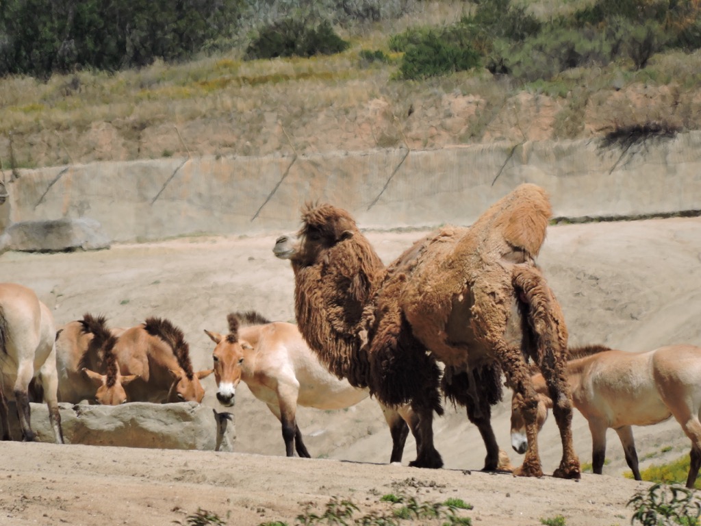 A big part of understanding paleontology is to understand the way things work today. In interpreting fossils, paleontologists first look to the modern world for analogs, whether it's identifying a species, interpreting behavior and ecological relationships, or even understanding how a fossil came to be preserved at all – the subbranch of paleontology known as taphonomy.A few weeks ago Brett and I were visiting the San Diego Zoo's Safari Park, located in Escondido about 45 minutes from the museum. While photographing Przewalski's horses and Bactrian camels (above, both of which have Pleistocene relatives preserved at Diamond Valley Lake), Brett noticed an odd shape located on a hillside in the exhibit (inside the red circle below):
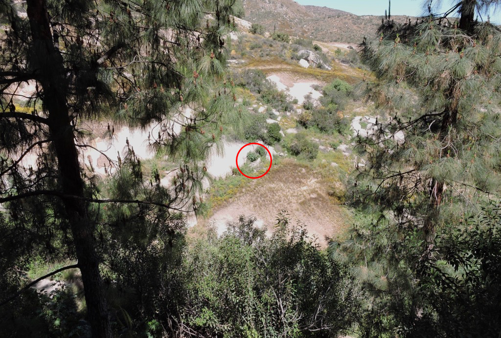 Fortunately I have a camera with a pretty powerful zoom lens, so I was able to get a closeup look at the area:
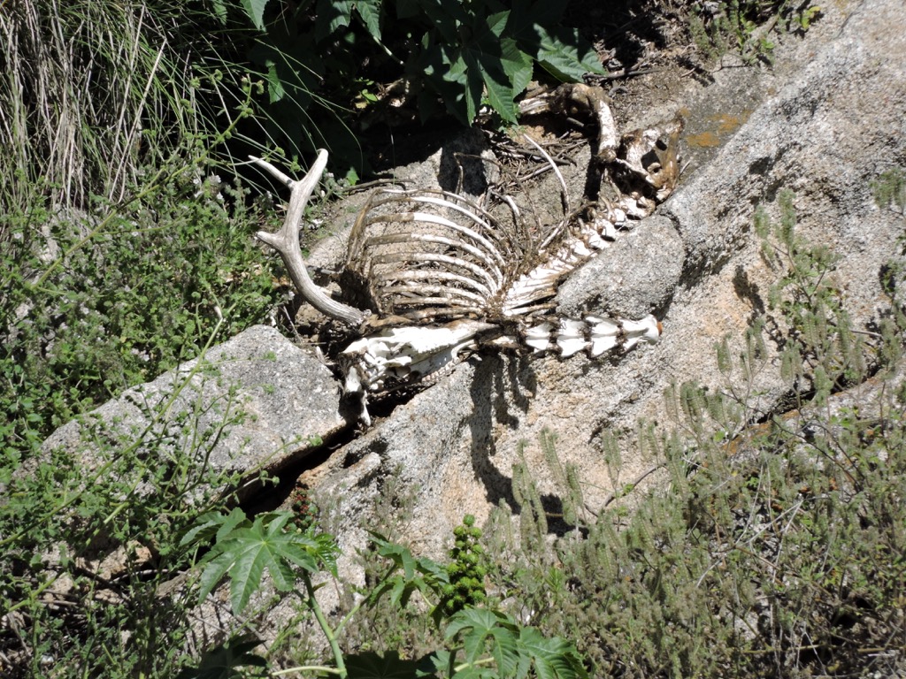 A skeleton! There's enough here to make an attempt at some identifications:
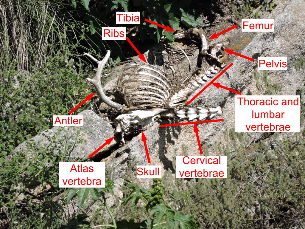 Quite a lot of this skeleton is present, and we can say a fair amount about it. First, the presence of an antler is significant. We can tell it's an antler and not a horn from the multiple tines (at least three), and because it has a growth pedicle. That puts this in the Cervidae, the deer family. Unless it's an exotic animal from the zoo collection or some other source (unlikely) that means a mule deer, Odocoileus hemionus.The skeleton is mostly intact and articulated, and there are still a few bits and pieces of skin and fur attached in places. The neck has bent backwards, a common occurrence after death. As the carcass dries out, the nuchal ligament that runs along the back of the neck tends to shorten, pulling the head backwards over the back.There has been some disturbance to the skeleton. The head has been pulled off the body. With the attached atlas vertebra, it has been flipped upside down (relative to the body) and rotated 180 degrees so that the snout is facing the back. The lower jaw is missing entirely. The lower parts of the hind limbs are probably obscured by vegetation, but the front limbs including the shoulder blades are missing. With the skeleton sitting on a steep hillside, we wondered if some the bones had ended up downhill, and we spent a half hour or so with the camera examining the hillside, with some success. About 10-20 m down the hill we located an ulna:
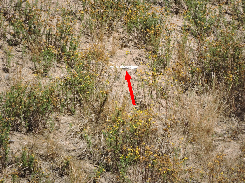 ... a partial scapula:
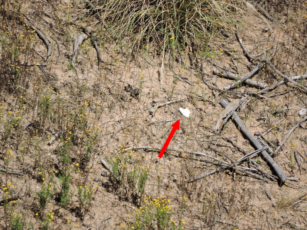 ...and a humerus:
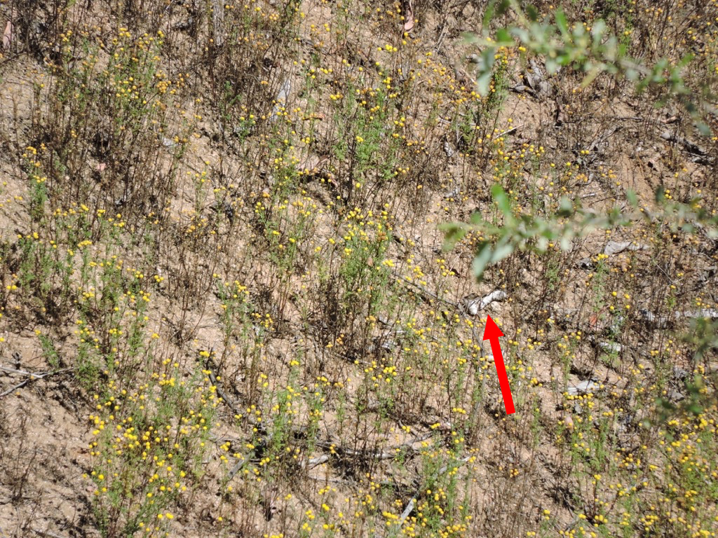 We never did find the lower jaw.So, what's the story of this skeleton? I'm not sure why the deer was in the enclosure in the first place, but there are steep cliffs in the area; I suppose it's possible the deer could have fallen in, and maybe even been killed in the fall. There was some disturbance of the carcass after death, but probably not by really large scavengers. I assume zoo staff would have removed the carcass or left it alone entirely, so they probably aren't responsible for the disturbance. I also think it's unlikely that large carnivorans such as coyotes, black bears, and mountain lions are allowed to roam freely with the zoo's herbivores, and they likely would have torn up the skeleton more than what we see. Small carnivores are a different matter. On the same trip we saw squirrels passing back and forth through small openings in some of the fences; a feral cat could have passed through the same openings. For that matter, squirrels and other rodents will also scavenge carcasses, mostly looking for calcium in the bones. But the most likely culprits are scavenging birds such as ravens, crows, and turkey vultures. These birds are large enough to strip a carcass and even remove some pieces (like the skull and the front legs), but often leave large parts of the skeleton intact, and, of course, they have no difficulty in gaining access to the enclosure.Since this skeleton is in a zoo, it's somewhat protected from the full range of taphonomic processes it would be exposed to in the wild, such as large mammalian scavengers, but that alone isn't enough to save it. Over time, the skeleton will break down in the sun. The outer cortical bone will deteriorate and flake away, and once the interior bone is exposed the breakdown will occur even faster. Occasional rainstorms will speed this process, as will insects and rodents feeding on the bone itself. More robust fragments, such as the ends of limb bones and flakes of tooth enamel may survive a bit longer and wash down the hill. There is a small stream at the bottom of the hill, so it's conceivable (if unlikely) that a few of these more robust fragments could be buried in stream sediments. Within a few years, if anything of this deer survives, that will probably be all there is.
Fossil Friday-horse skull fragments
 Vertebrate paleontologists rarely get to examine whole skeletons (really, almost never). Even individual bones or regions such as skulls are usually fragments, often broken in odd ways, and identification can be a challenge. That's why paleontologists will keep around lots of well-illustrated references, reference collections of modern animal skeletons, and increasingly, 3D images of identified specimens. A lot of identification work is just comparing your unknown specimen to known ones and finding a match.For the last several weeks WSC volunteer Nathan Bonde has been preparing a block of sediment that came to the museum through a mitigation project (unfortunately with rather limited data). The sediment had several bone and possible tooth fragments sticking out, but it wasn't immediately clear what the material was or if it all belonged to the same animal.
 It quickly became apparent that the tooth was from a horse, apparently a upper left 3rd premolar or 1st molar (shown below next to a zebra skull from Brett's collection):
 The bone fragments were a little tougher. Continued cleaning showed that most of the bone joined together into a single large fragment, shown at the top of the page. The size and overall complexity suggested a skull fragment, which was bolstered by the presence of a bony tube that looked like the external auditory meatus, part of the ear structure. Since we knew that a horse was a strong possibility (based on the tooth), we compared the fragment to the zebra skull:
 If you're having trouble seeing how these match up, here's an annotated version:
 The area outlined in red on the zebra skull is the approximate preserved part of the fossil; note that the fossil is quite a bit larger than the modern specimen. The numbered features are:
- Right occipital condyle (the articulation with the 1st neck vertebra)
- Condyloid fossa
- Jugular process
- External auditory meatus
- Glenoid fossa (the articulation point for the lower jaw)
- Temporal fossa (opening for the muscles that close the lower jaw)
- Zygomatic process of the squamosal (cheekbone)
Here's another view, oblique and from behind, showing more clearly how the zygomatic process projects out, forming the lateral side of the temporal fossa: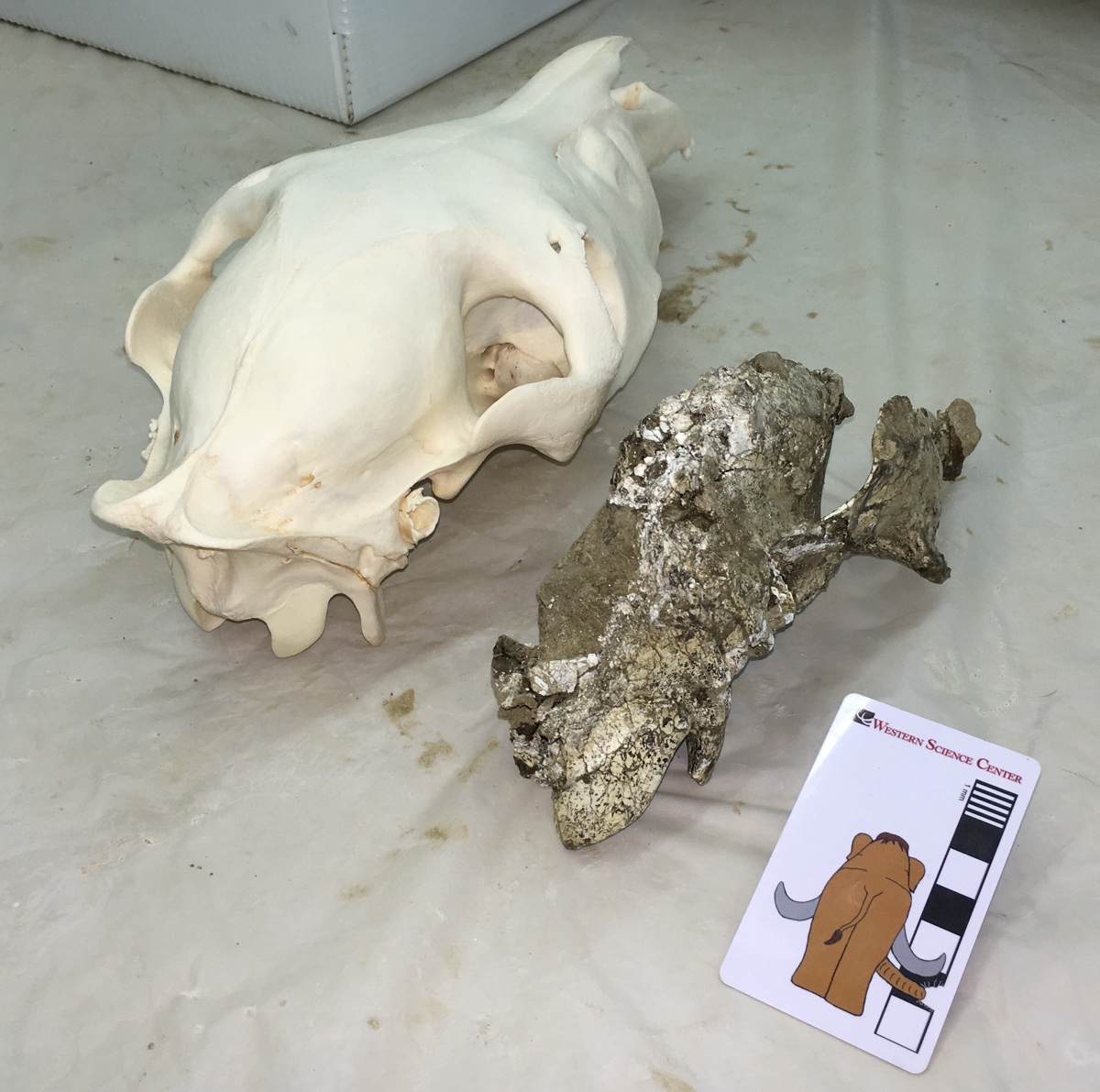 Tooth size is apparently not a reliable indicator of body size in horses, and our modern specimen is both sub-adult and a relatively small animal. Nevertheless, in this instance we seem to be dealing with a fairly large horse; both the tooth and the skull fragment are much larger than our reference skull. If you look closely at the images above, you may also notice differences in the shape of the enamel ridges on the teeth, and somewhat different shapes and orientations in some of the cranial bones. While both these skulls are from the genus Equus, they do not represent the same species, which isn't really surprising considering that they're separated by at least 10,000 kilometers and at least 20,000 years.
Fossil Friday-mastodon jaw fragments
 We're entering the final hours of our crowdfunding campaign at experiment.com, and are very close to our funding goal. This week we're featuring another specimen that will be included in this study.While the bulk of the Western Science Center's collection came from Diamond Valley Lake, we are a regional repository with a number of collections from other localities. This specimen was recovered during a mitigation project in Temecula in southwestern Riverside County, and after reconstruction by WSC volunteers it became apparent we had a significant portion of a mastodon lower jaw. The image is in dorsal view, with anterior to the left. Parts of both dentaries are preserved, although there is considerably more of the left dentary present.Portions of four teeth are preserved, the lower second and third molars. On the left side the second molar is complete, and the third molar is nearly so, allowing us to get length and width measurements for inclusion in our project; it turns out that these teeth are long and narrow, exactly what we've come to expect from California mastodons. The second molars are pretty heavily worn, while the third molars show wear only on the first two lophs. That is very close to the same wear state found in Max, so these two were probably close to the same age.If you haven't already done so, please go to experiment.com/mastodon and donate to our project, so we can start to understand how mastodons were distributed through North America.
Fossil Friday - Bison latifrons skull
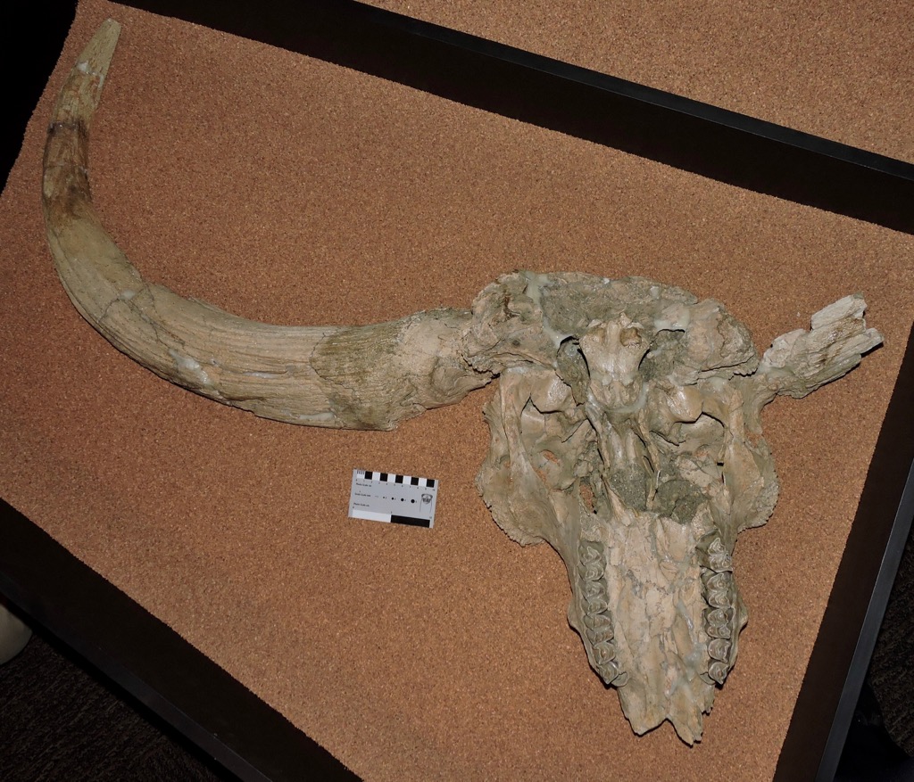 We still have a few days left in our crowdfunding campaign to study mastodons, but for Fossil Friday this week I'm stepping away from mastodons to look at a different animal. Earlier this week the U. S. Government passed a law designating the American bison (Bison bison) as the United States' National Mammal. Bison are prominent in the fossil record, including at Diamond Valley Lake.As I've mentioned in previous posts, there were two species of bison present at Diamond Valley Lake. The more common species was Bison antiquus, a possible direct ancestor to the modern B. bison. The rarer, larger species was the long-horned bison, B. latifrons.Diamond Valley Lake's best B. latifrons skull, shown above, is on permanent exhibit at the museum. It's displayed in ventral view, and in the image the front of the skull is at the bottom. The premaxillae forming the tip of the upper jaws is missing, as is the right horn core, but otherwise the skull is mostly complete if a bit deformed. One thing to keep in mind when looking at fossils of Bison latifrons (and, indeed, any fossil bovid) is that the preserved horn is simply the bone core of the horn. In life the core is covered with a keratin sheath, making the living horn considerably larger.It's somehow fitting that the United States' national mammal is in fact an immigrant to North America, and a relatively recent one at that. Bison were one of the last large mammals from the old world to spread across North America (some of the other large Old World immigrants, such as deer, mammoths, and mastodons, and South American ground sloths, all arrived earlier). In fact, bison only made it south of the 55th parallel (which runs through central Canada) roughly 250,000 years ago, and then rapidly spread across southern Canada and the United States. The first appearance of bison south of the 55th parallel is used by paleontologists at the marker for the start of the Rancholabrean North American Land Mammal Age (NALMA), the last NALMA of the Ice Age.During the Rancholabrean, various bison species ranged across the entire northern hemisphere. While some bison species went extinct at the end of the Ice Age, the genus Bison survived and thrived in the Holocene in both Eurasia and especially in North America. Two extant species are recognized, the American bison (Bison bison) (seen at Yellowstone National Park) and the European bison (Bison bonasus) (seen at the San Diego Zoo Safari Park):
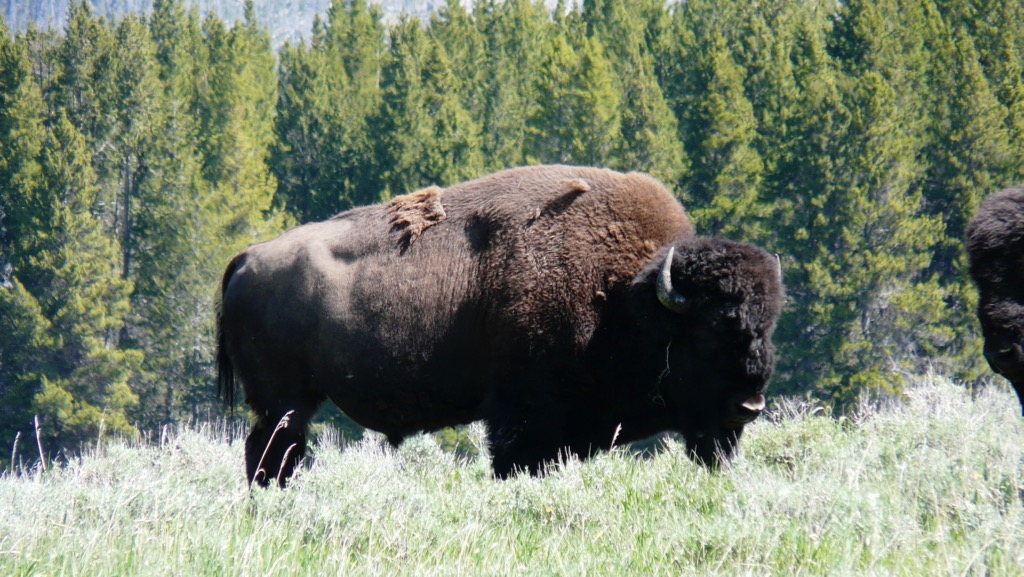
 This wasn't to last, as bison were almost completely wiped out by overhunting and habitat loss over the last few hundred years. Both species have been protected and are slowly recovering, although populations were reduced to such a low number in the past that genetic diversity is quite low. Even so, there are once again wild herds of both species living in protected areas.
Fossil Friday - Old Man Mastodon
 Closing our our Month of Mastodons is one of the more complete mastodon skulls from Diamond Valley Lake, a specimen that has several interesting features in addition to its relatively good preservation.First, to explain what you're looking at, here's an annotated version:
 The skull is laying on its right side, at a slight angle. Much of the braincase seems to be missing, although this area hasn't been fully prepared, so there could be more there than there appears. The left dentary (lower jaw) is present, but disarticulated; it's not clear if the right dentary is still in the bottom of the jacket.So, what can we say about this skull? Well, for starters it looks pretty large, with a massive tusk. I haven't measured it out yet, but I suspect that based on size this will probably be a male mastodon. Zooming in on the teeth gives us more information:
 Both upper 3rd molars are visible, as is the lower left 3rd molar. These teeth are in wear across their entire occlusal surfaces. Also notice the flat area outlined in red below, located in front of the upper left 3rd molar:
 This is the closed-up socket for the 2nd molar, which has completely worn down and fallen out. With the 2nd molars gone and the 3rd molars heavily worn, this was a pretty old mastodon, most likely over 50 years old. He was much older than Max, who still had his 2nd molars, and in fact is the oldest mastodon I've seen from Diamond Valley Lake so far.There is another interesting feature in these teeth, specifically in the lower 3rd molar. The red line below traces the outside edge of the chewing surface of the tooth (the "labial edge of the occlusal surface"):
 The tooth is most heavily worn in the middle, and is higher both in the front and the back. This is the only time I've seen this wear pattern in a mastodon tooth, and I'm not sure how it formed. Here's the issue: like other advanced proboscideans, mastodons replaced their teeth horizontally. That means the front of the tooth erupts first. Because of this, at any given point in time after eruption, the more anterior parts of the tooth have been functional for longer than the more posterior parts. Because of this there should be a wear gradient, with the tooth being progressively less worn as you move from the front of the tooth to the back. And normally that's exactly what we see in mastodons, mammoths, and other proboscideans with horizontal tooth replacement. So how did this tooth get most heavily worn in the middle? Was the tooth injured or deformed in some way? Did the mastodon's diet change at some point, changing the wear rate? Did the mastodon suffer an injury to his jaw or jaw musculature that affected his ability to chew, altering the occlusion pattern of his teeth? (Max had multiple injuries to his lower jaw, so this type of injury may be common in male mastodons.) Or is there something else going on?This mastodon skull is currently on display in the "Stories from Bones" exhibit at WSC, but if you haven't seen it yet you'll have to hurry; "Stories from Bones" closes on May 29.With complete 3rd molars, this mastodon is also one of the specimens that is contributing data to our mastodon tooth research project, "Mastodons of Unusual Size". We only have a few days left in our crowdfunding campaign to support this research, and we're still well short of our goal, so donate today and help us understand mastodons a little better!
Fossil Friday - partial mastodon skull
 As we continue our month of mastodons in support of our crowdfunding campaign at experiment.com, this week we have a partial mastodon skull from Diamond Valley Lake.When I say partial, I mean very partial. Practically all that's left of this specimen are the upper teeth and one tusk. Almost all the bone is missing, a somewhat odd preservation type that we get occasionally at Diamond Valley Lake. We haven't taken measurements on the tusk, but it seems to be massive, suggesting that this might be a male. The skull is laying upside down, and oriented so that anterior is toward the camera (this large jacket was sitting on a storage shelf, and would have been difficult to move quickly to get Fossil Friday photos in other orientations). The two upper third molars are visible, but there's no preserved trace of the second molars. The crowns of the third molars are well preserved, so this is a specimen we'll be able to include in our study. Every loph on the molars is in wear, with the last lophs just starting to wear, so this animal was presumably a bit older than Max (who still had second molars and unworn last lophs on the the third molars).We have just under two weeks to go in our crowdfunding campaign to gather data on mastodon tooth shape from localities across the country, and we still have a long way to go, so please visit experiment.com/mastodon and give what you can to help us with this study.
Fossil Friday - skull of "Little Stevie" the mastodon
 As promised, during our crowdfunding campaign to work on mastodons I'm featuring mastodons in all my Fossil Friday posts. This week is the partial skull of "Little Stevie" from the WSC collections. "Little Stevie" was the most complete mastodon found at Diamond Valley Lake, with about 40% of the skeleton recovered. Thanks to a donation by Eric and Gisela Gosch, most of "Little Stevie's" skeleton is on display in a case embedded in the floor of the museum, but the partially prepared skull is in the collections. Below is a marked-up version of the image above:
 The skull is laying upside down, and the yellow arrow is pointing anteriorly. That means you're looking at the right side of the skull. The 2nd and 3rd upper molars are indicated in blue, and the right occipital condyle is labeled (the skull's articulation with the first neck vertebra). The broken cheek bones are visible close to the camera, as are the internal choanae (the internal openings for the nostrils) just behind the 3rd molar. There are also a number of postcranial bones in the jacket, including several ribs (there's one below the yellow arrow) and two thoracic vertebrae indicated by the red arrows. Both thoracic vertebrae are missing their epiphyses, indicating that "Little Stevie" was still growing. However, the femur associated with this skeleton has fused epiphyses, and at least the first two lophs on the third molar are in wear, which suggests that "Little Stevie" was close to full grown, maybe early to mid-20s.We don't know for sure if "Little Stevie" is a male or a female. The best markers for determining sex in a mastodon are the pelvis and the tusks, neither of which is well preserved in "Little Stevie" (although we may eventually be able to get a pelvic measurement). "Little Stevie's" femur is very close to the size and proportions of the Java Site mastodon from New York, which is thought to be a female, but that's not a very reliable indicator of sex."Little Stevie" does include both 2nd and 3rd molars and a femur, all of which are bones we're examining in our mastodon study, so that means "Little Stevie" is included in our project. Help us collect more data to compare to "Little Stevie" by donating at experiment.com/mastodon.
Fossil Friday - mastodon molar
 On Monday Eric Scott and I launched a crowdfunding campaign to support a mastodon research project we've been working on. We believe California (or maybe western) mastodons have different tooth proportions than mastodons from other part of the country, and in order to test that hypothesis we need to travel to other museums to collect measurements on mastodon specimens from as many locations as possible. In recognition of that campaign, for the next month all my Fossil Friday posts will feature mastodons.Above is a left upper 3rd molar from Diamond Valley Lake, shown in occlusal view. This tooth was found shattered and painstakingly reconstructed, so even though it's incomplete it's an impressive specimen. Below is a lingual view of the same tooth:
 The anterior loph (the large ridge on the crown of the tooth) is mostly missing on this specimen, but the other lophs show little or no wear, so this tooth was either unerupted or had only recently erupted. As an aside, while I haven't checked the numbers to confirm this, a lot of the Diamond Valley Lake mastodons seem to have died when the third molars were partially erupted, maybe in their mid-20s to mid-30s.Since the front of this tooth is missing we can't take a reliable measurement of its length, which means this specimen will probably not end up in our project database; we need to be able to compare the length and width of the tooth crown. But even incomplete, this seems to be a remarkably small tooth even by California standards. Interestingly, our data so far suggests that molar size is not a very good indicator of body size (Max was a big animal with relatively small molars), so even though this tooth is small it doesn't mean it came from a small mastodon.We hope you'll visit experiment.com to help us study these mastodons!