

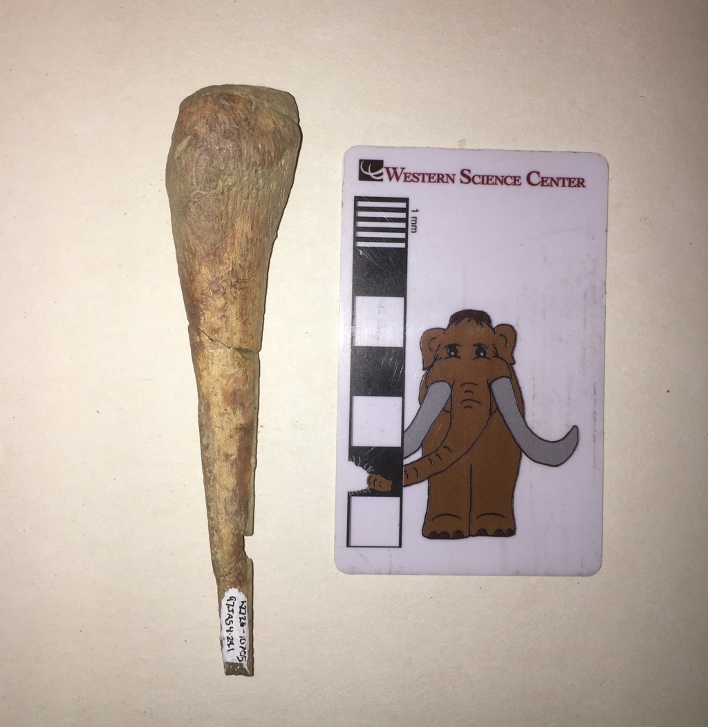
 One of the joys of paleontology is that every fossil has a story. Through our understanding of anatomy, geology, ecology, and a host of other field, we can often reveal part of that story, and even a relatively small, nondescript fossil takes on a larger meaning.The small, pointed bone shown above is the right fourth metacarpal from a horse (Equus). The metacarpals are the bones that make up the hand or forefoot. At the proximal end they articulate with the wrist bones (the carpals) and often with the adjacent metacarpals. At the distal end they normally articulate with the first finger bones (the phalanges). In humans, the fourth metacarpal articulates with the ring finger. Our fingers (or digits) are numbered from 1 to 5, with the thumb being Digit I and the pinkie finger being Digit V; thus the ring finger is Digit IV. Toes are numbered the same way, with the big toe as Digit I.Horses famously only have a single hoof on each foot, corresponding to a single finger (on the front feet) or toe (on the back feet). The distal end of the bone shown above (at the bottom of the photo) clearly does not have any kind of articulation for the bones that make up the single finger on the horse's right foot. So what's going on?In horses, the hand bone that supports the animal's weight is the third metacarpal, not the fourth one. The right third metacarpal (probably from the same individual) is shown below:
 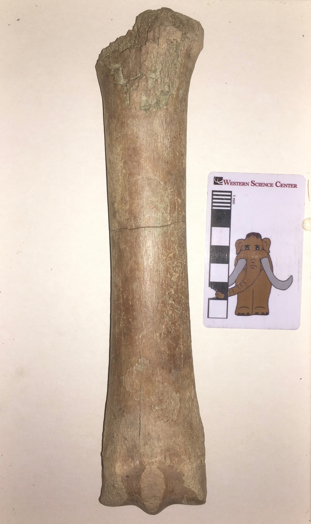 This massive bone is well-suited to holding up a horse's weight. So what about the fourth metacarpal? If it doesn't support a hoof, what does it do?As far as we can tell, pretty much nothing.The fourth metacarpal is a remnant of the horse's evolutionary history. While modern Equus is limited to a single hoof on each foot, supported by Digit III, horses' ancestors and extinct relatives had multiple hooves on each foot. The lateral toes were gradually lost, starting with Digits I and V, and later with Digits II and IV. The key here is gradually. Each digit eventually became small enough that it no longer functioned for walking; there would be a tiny hoof, but it didn't reach the ground. The hoof would eventually be lost, leaving only the metacarpal with no attached finger or toe. After even more time and generations, even the metacarpal is lost. But the whole process takes several million years.In the horse lineage, the first and fifth metacarpals were lost tens of millions of years ago. But the reduction of the second and fourth metacarpals has happened much more recently. Within the last 10 million years horse ancestors still had functional second and fourth toes, even if they were smaller than the central third toe. A few million years ago the second and fourth toes were finally lost altogether, leaving their corresponding metacarpals and metatarsals as the last remaining remnants of these digits. And that's where horses are today. The second and fourth metacarpals (and metatarsals) have lost their toes, and even the articulation to attach to the toes, and they're greatly reduced in size. At the proximal end they still articulate with the wrist bones and the third metacarpal (see the posterior view below). The presence of the fourth metacarpal is evolution caught in the act, a snapshot of a dynamic process that seems static because it happens so slowly. If horses survive a few million more years, the fourth metacarpal will probably be entirely lost, so that there is only a small slice of time (geologically speaking) when such a fossil could be preserved.
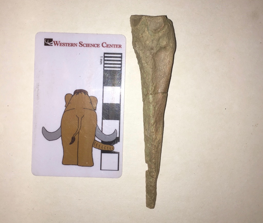
Fossil Friday - Paramylodon skull
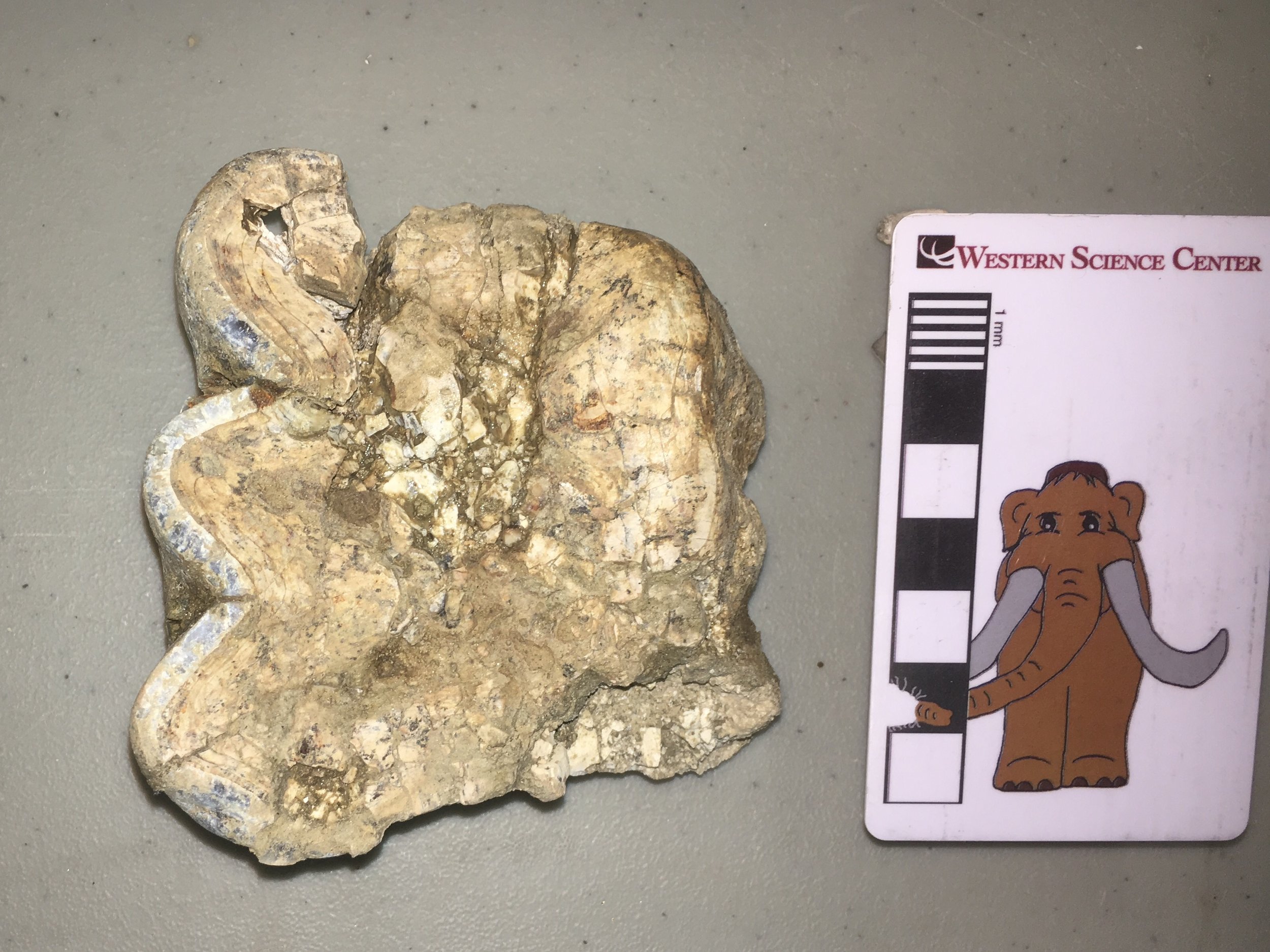
 Even if a bone is lucky enough to be preserved as a fossil, time is not always kind. There are numerous ways a bone can be altered after burial, including being smushed by the weight of overlying sediment.The lump of bone shown above is actually a nearly complete skull of the ground sloth Paramylodon harlani in dorsal view, collected from the El Casco substation in northern Riverside County. It might be a little difficult to interpret because it has been deformed post-burial. Below is an annotated version:
 The skull is pushed over so that the midline is displaced to the left. This makes features from the right side like the right eye socket easily visible in dorsal view, while hiding the same features from the left side. The entire skull is also flattened, resulting in the wide foramen magnum at the back of the skull.Here's the ventral view:
 And the annotated version:
 Much of the basicranial part of the skull is a big-ole' indecipherable mess, but there are a lot of identifiable features, including all of the tooth sockets.While there are other Paramylodon remains from El Casco, this is the only sloth skull recovered from the site. Even deformed specimens can be significant!
Fossil Friday - tree frog humerus
 The majority of fossils from the Diamond Valley Lake deposits are from small animals. While small mammals are most common, there are substantial numbers of birds, reptiles, and amphibians.The bone shown above is a portion of a frog's left humerus, seen in ventral view; the bars behind it are 1 mm wide. Approximately the distal third of the bone is preserved, with the distal end (the elbow joint) on the right. The expanded distal end is divided into two parts; the larger, more spherical portion is the articular condyle, while the smaller medial portion (on the lower right) is the medial epicondyle.This humerus has been identified as Pseudacris sp.. Pseudacris is a widespread North American frog genus that includes various tree frogs, chorus frogs, and peepers, with several species still living in the southwestern US and Mexico. Below is an eastern example, a spring peeper (Pseudacris crucifer):
 An interesting feature of many frog species is that the humeri are sexually dimorphic. Males have a large flange, the crista medialis, that extends proximally from the medial epicondyle and make the humerus look much wider in ventral view. This specimen lacks a large crista medialis, so if that is a characteristic on Pseudacris it suggests this specimen is a female.
Fossil Friday - Bison atlas vertebra, part 2
 This week's Fossil Friday features one of Diamond Valley Lake's most common large mammals, the bison.Specifically, this bone is the atlas vertebra, the first vertebra in the neck. It's shown above in anterior view. The large hole in the bone is the neural canal, which carries the spinal cord in the live animal. The large concavities on each side of the neural canal are the articulations for the skull's occipital condyles.Below is the posterior view:
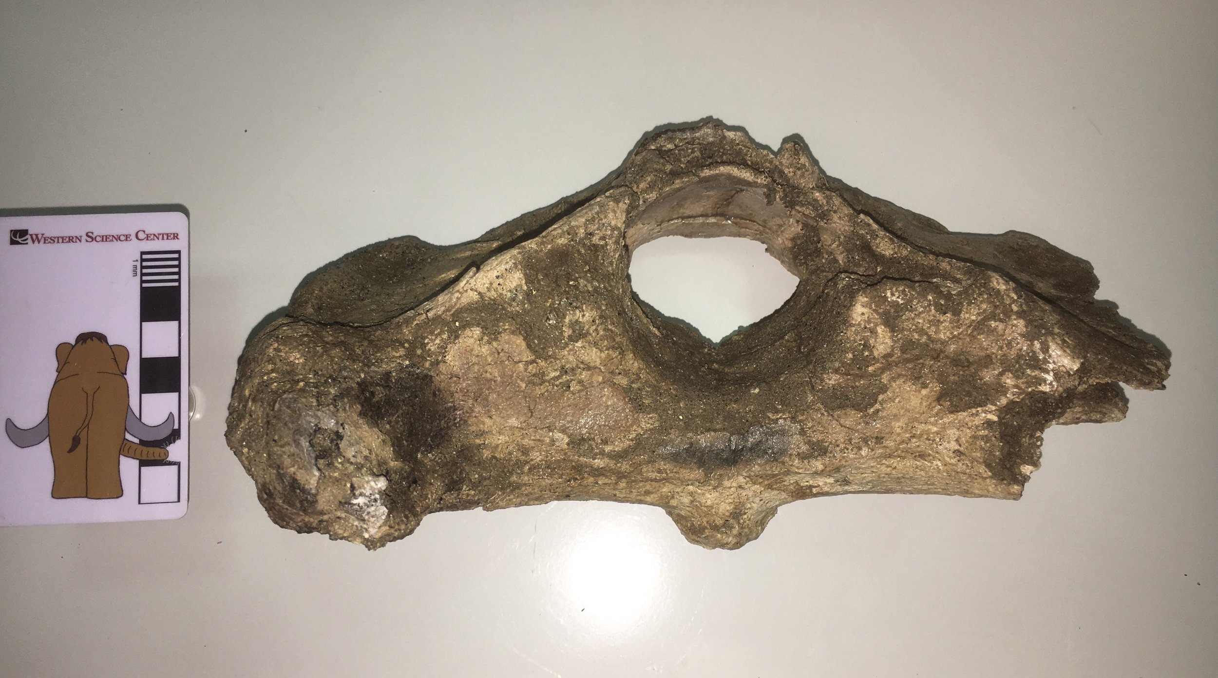 The preservation is a little rougher on this side. The posterior edge of the neural arch (the top of the neural canal) is missing, as is most of the right transverse process. The flat areas to each side of and slightly below the neural canal are the articular surfaces for the 2nd cervical vertebra, the axis.Here is the dorsal view:
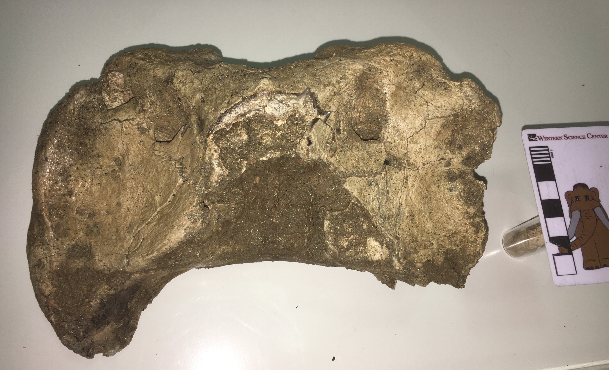 ...and the ventral view:
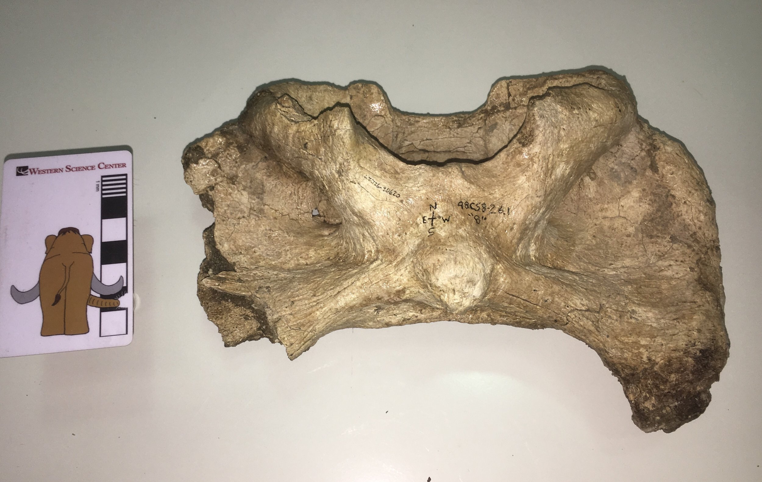 This vertebra is identified in our records as Bison antiquus. It comes from the West Dam area of the lake; sediments there are mostly less than 20,000 years old and seem to only produce Bison antiquus and not the large Bison latifrons. Bison antiquus is generally considered to be closely related or ancestral to the modern Bison bison, and indeed this atlas looks very similar to a modern bison's. This make an interesting contrast to the DVL bison atlas I wrote about back in May 2015. That vertebra was identified as Bison sp., but its shape is a bit different from this one (I even commented at the time that it differed from Bison bison). So perhaps our two extinct bison species really do have distinct atlas shapes. At some point we'll have to compare these bones to atlases that are associated with skulls to confirm that we're seeing specific differences and not individual or sexual variation.
Fossil Friday - worn mastodon tooth
 Volunteer Joe Reavis been hard at work on a collection of fossils from a mitigation project in Murrieta that includes a lot of mastodon material. As far as we can tell so far, all of the mastodon material is consistent with one individual, although we did confirm yesterday that there is non-mastodon material in the same collection.The specimen shown above is a molar from a mastodon. It's in pretty rough shape, but I think it's the upper right second molar. If that's correct, then the occlusal view above has anterior at the top, and the lateral side of the tooth is on the left. The three loops of enamel on the left are the remnants of three lophs, which indicates that this is either a 4th premolar, 1st molar, or 2nd molar (the 2nd and 3rd premolars only have 2 lophs, and the 3rd molar has 4 or 5 lophs). The length of the tooth is comparable to other 2nd molars in our collection.Upper teeth in mastodons wear more rapidly on the medial side than the lateral side. If we look at this tooth in lateral view, we can see that there is a only about 2 cm of enamel remaining; the rest of the lophs have been worn away:
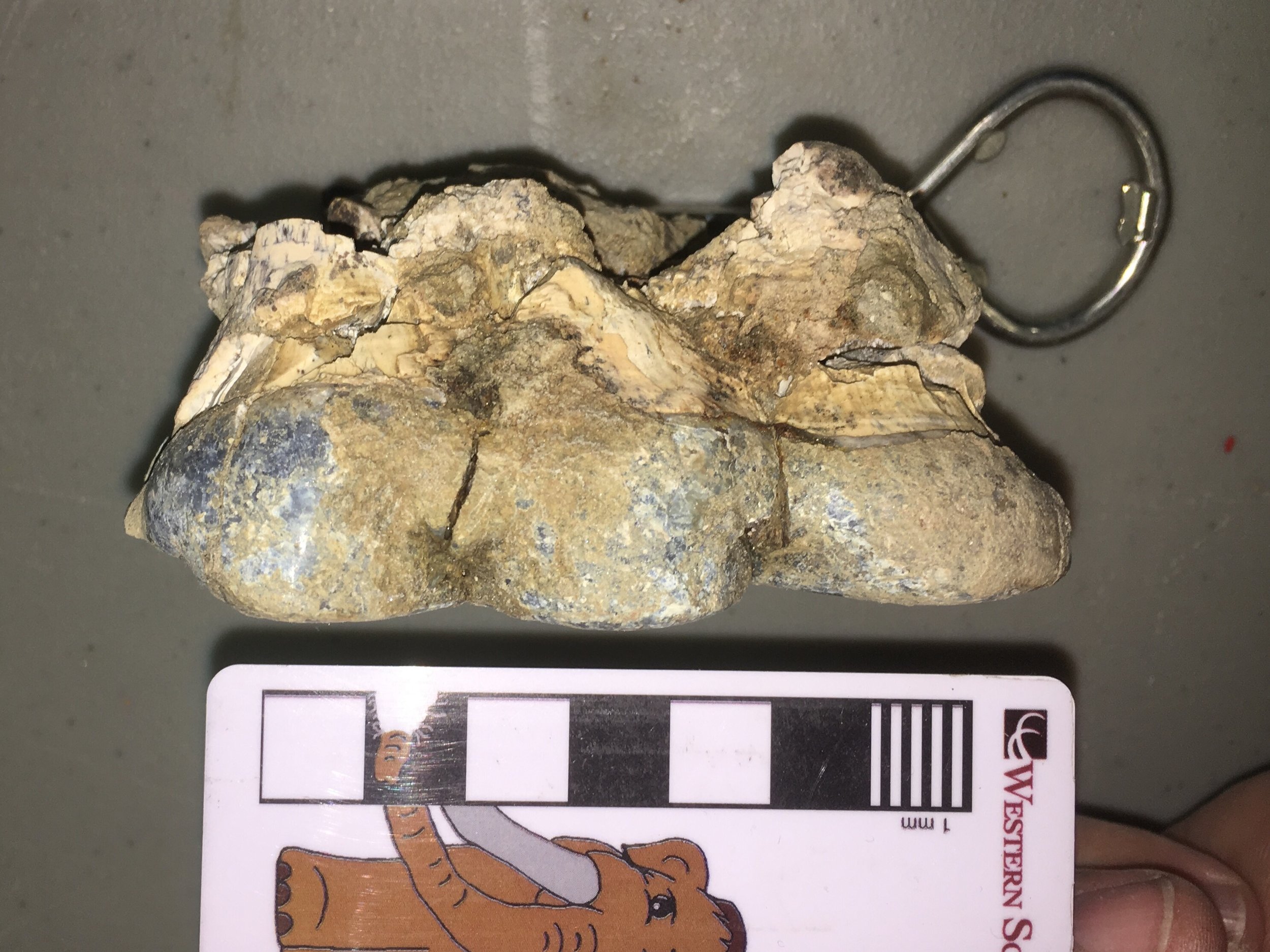 On the medial side, the wear is even more dramatic:
 If you're having a hard time telling what's going on here, it's because the enamel is completely gone. The tooth is worn all the way down into the tops of the roots.In most animals this would indicate that the animal was very old, but the horizontal tooth replacement in mastodons and most other proboscideans changes the equation. The 2nd molar typically wears away and falls out sometime around age 40 (very approximately). This tooth still had its roots (they were broken post-mortem), so this mastodon was probably around 40 years old when it died.
Fossil Friday - chewed-up Bison tibia
 I recently finished reading Anthony Martin's book about dinosaur trace fossils, Dinosaurs Without Bones, so I've had trace fossils on my mind. Even though I'm not a trace fossil specialist I find them intriguing, because they are essentially fossilized behavior. The bone shown here is the distal part of a right tibia from a bison (it's from the older end of the valley, so it could be either Bison antiquus or Bison latifrons). Above is the anterior view, with the bottom of the bone (at the ankle joint) on the left. The proximal part, including the knee joint, is missing. Below is the posterior view of the same bone:
 Even in these views, you may have noticed that the distal end of the bone is a little misshapen. Close-ups reveal that this bone is absolutely riddled with bite marks, presumably from a predator/scavenger gnawing on the bone:


 The broken proximal end also has plenty of bite marks:
 This bone is crying out for a more detailed study. There appear to be at least 2-3 different sets of scratches with different widths. Does this indicate that there were different-sized scavengers? If so, are we looking at different species taking turns (maybe dire wolves followed by coyotes), or different ages of the same species (adult wolves and their pups)? If the scratches show cross-cutting relationships, it might be possible to figure out if the big animals were eating before the small ones, or the other way around, or at the same time.
Fossil Friday - sloth mandible
 Greg McDonald's visit last month to look at sloth remains gave us a reason to open our display cases, which include some of our best sloth fossils.One of the exhibit specimens is a nearly complete mandible of Paramylodon harlani, the most common sloth from Diamond Valley Lake. The image is a dorsal view, with anterior to the right. The mandible is nearly complete, if a bit crushed. It's missing the articulation on the right side, part of the anterior end, and all the teeth, but is otherwise in good shape.Paramylodon lower jaws are scoop-shaped and toothless at the tip. This is very different from their distant relative Megalonyx, that has a pair of huge chisel-like teeth at the tip. Even though the teeth are missing, the shape of the sockets also reveals a different cross-section that the more rectangular teeth of Megalonyx. For additional examples of Paramylodon jaws, see our earlier Fossil Friday posts here and here.---This weekend I'll be at the Western Association of Vertebrate Paleontology meeting at Yavapai College in Arizona, presenting on the Mastodons of Unusual Size project and on the Stepping Out of the Past exhibit. Follow @MaxMastodon on Twitter for updates.
Fossil Friday - camel lumbar vertebra
 While we only have one well-preserved skull of the extinct camel Camelops hesternus from Diamond Valley Lake, we have a large number of post-cranial remains.The bone shown above is a lumbar vertebra, seen in anterior view. This seems to be the 7th lumbar, the last one in the series before the sacrum (which was also recovered from this individual, along with several other bones). The prominent curved structures above and on each side of the neural canal are the prezygopophyses. These articulated with the postzygophyses of the 6th lumbar. The strong curvature would largely lock the two vertebrae together, resulting in a relatively inflexible lumbar region.Here is the posterior view, with the postzygophyses visible:
 And the left lateral view:
 The neural spine is broken on this specimen, as are the transverse processes (the right one is missing entirely). The broken surfaces are packed with sediment and abraded, indicating that they were broken off before burial. That is interesting considering that there are multiple associated bones with this specimen; that makes it less likely that the bone was damaged by, say, washing down a river. Could this damage have been caused by scavenging? I tried looking at the bone with low-angle light, which sometimes helps reveal bite marks on the surface:
 Sure enough, these are apparent bite marks on the bottom edge of the centrum. Below is the preserved part of the left transverse process, with apparent bits marks along its entire length:
 There are other possible bite marks scattered across this vertebra. Moreover, close examination also revealed possible insect feeding traces in various places, including on one of the prezygapophyses:
 These possible traces seem to be extremely common on bones from Diamond Valley Lake, possibly occurring on half or more of the large specimens. Clearly, besides studying the bones themselves, there's a lot of potential in the DVL trace fossils as well.
Fossils of my youth
 Inspired by the #GatewayFossil hashtag on Twitter, I'm reposting this piece that I originally published at "Updates from the Paleontology Lab" on June 9, 2009.My first exposure to fossils in the field (as opposed to in a museum) occurred when I was around 5 years old.When my father was a teenager he used to hunt with my grandfather in the mountains along the Botetourt-Craig County line in western Virginia, where he had casually noticed some fossils in the stream gravels (I’m not sure he knew at the time what they were). My mother has always had an interest in rocks and fossils (due to the influence of her grandfather), so my dad used to take us up to that area to look for fossils. We mostly found molds of crinoid column segments and occasional brachiopods in a brown sandstone (above). With the limited resources available to a relatively poor child growing up in the country at that time (no internet!), I didn’t know much more about those fossils, and doing my college work in Minnesota and Louisiana I never actually learned much about the rocks I collected in my youth, a point that was emphasized with the discovery last year of the Boxley stromatolite in Bedford County (where I grew up).Now that I’m back in Virginia with years of geologic training behind me I can look at these rocks in a new light. Last Friday, my father and I spent the day driving around Botetourt and Craig Counties, looking at rocks. Near Webster, VA we stopped at a railroad trestle close to the Blue Ridge Fault, with deformed Cambrian Rome Formation exposed in the trestle foundation:
 The concrete used to build the foundation also had large chunks of Conococheague Formation embedded in it (this bridge is only a couple of miles from the Boxley Blue Ridge Quarry):
 Moving into Craig County, we followed Stone Coal Road into the mountains (although I’m using the term “road” in its broadest sense):
 We came across several roadcuts of interbedded shales and fine-grained sandstones:
 These rocks are from the Devonian Chemung Formation (apparently redescribed as the Foreknobs Formation). They are tilted nearly 90 degrees, which is especially clear where the rocks intersect with the road (the lines leading toward the truck):
 In a few places the Chemung was rippled:
 And eventually we found some examples of the fossil crinoid and brachiopod molds that are common in this unit:
 Returning to my father’s house in Botetourt County I took a look at the rocks exposed under his house and storage shed. The exposures are pretty limited:
 Even so, there were some interesting bits, including this contact between a cross-bedded sandstone and a limestone:
 According to the Geologic Map of Virginia the mountain where Dad’s house is located includes both the Rome and Elbrook Formations (both Cambrian); I think this rock may expose the contact between them (with the Elbrook being the lighter-colored limestone at the top).
Fossil Friday - mastodon molar
 For the last few weeks, volunteer Joe Reavis has been diligently reconstructing a box of tooth fragments that came to the museum several years ago via a mitigation project in Murrieta, California. It quickly became apparent that the fragments were mastodon, and it seems they all come from a single tooth.The tooth is shown above in occlusal view, and the presence of four lophs show that it's a third molar. The angle of the lophs and comparison to other specimens in the WSC collection indicate that it's the upper left third molar.Here's the labial view:
 And the lingual view:
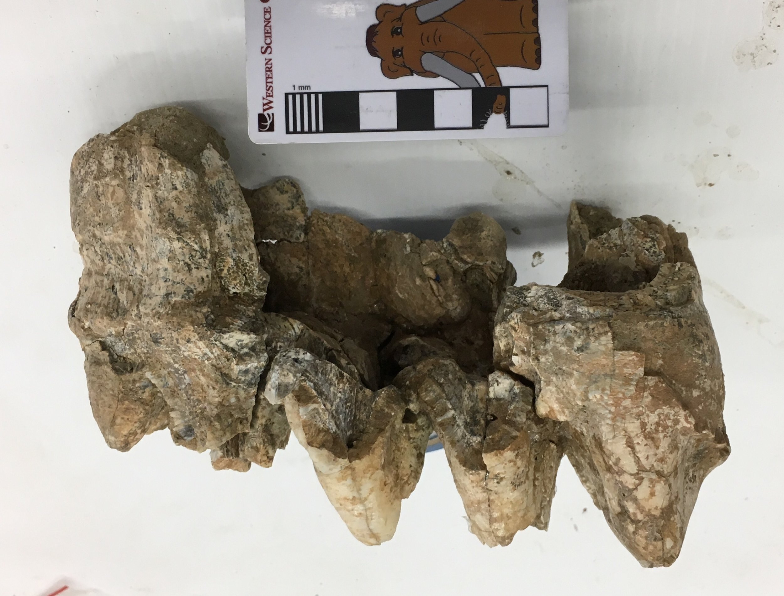 Looking past all the post-burial fracturing, this tooth shows no signs of any occlusal wear, and had probably not erupted. There is also enough preserved to get measurements for our "Mastodons of Unusual Size" project; the length:width ratio of this tooth is in the typical range for California specimens.The museum obtained several boxes of material from this site in Murrieta, but unfortunately it came with very little data or context. There is a fairly large amount of mastodon material included, including partial lower jaws with teeth and several limb elements, that are all consistent with a single individual. We're still in the process of preparing some of the other material.
Fossil Friday - oreodont skull
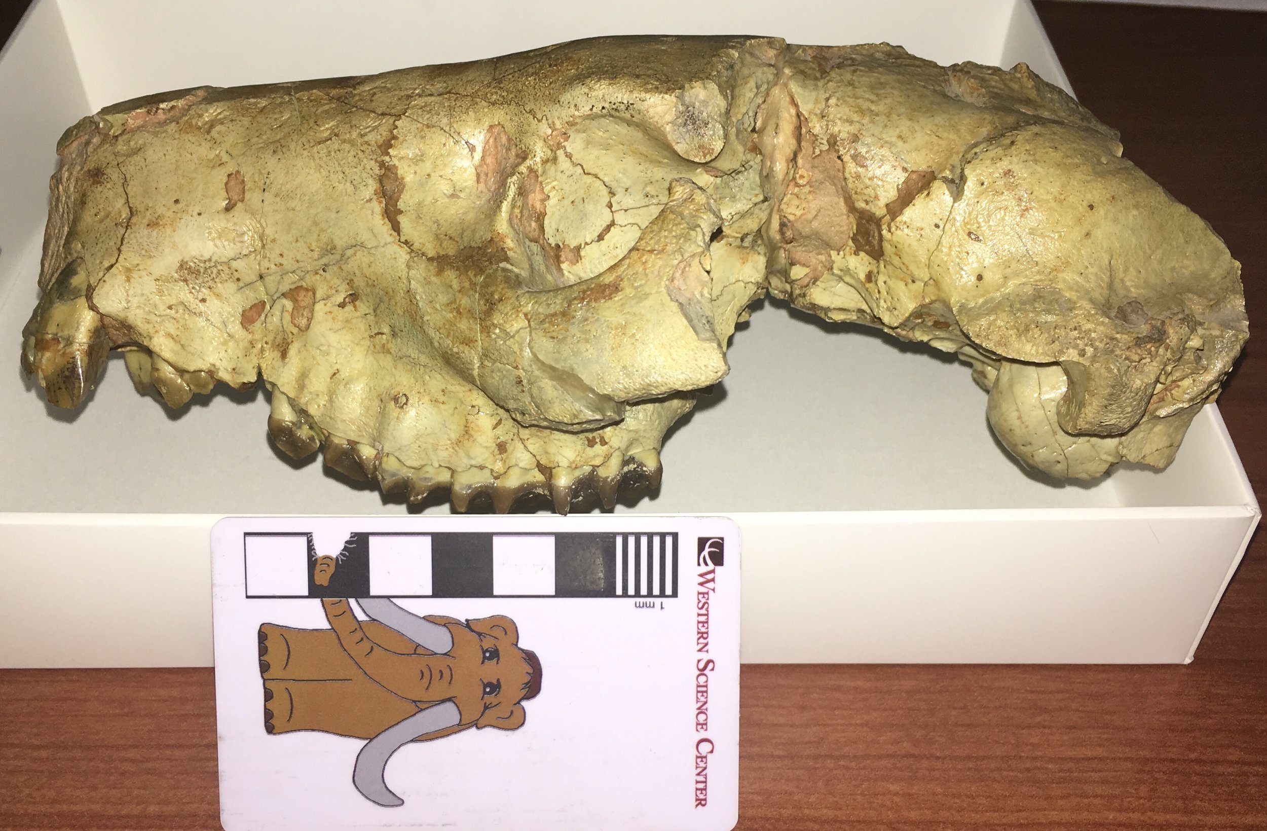 We're in the process of taking in a number of specimens collected by the late Harley Garbani, which are being donated to the museum by his wife Mary. The first item to come to us was a nicely preserved and prepared skull of an oreodont, the first in the WSC collection.Oreodonts are an extinct group of artiodactyls, generally thought to be distantly related to camels. They were very diverse and widespread in North America during the Oligocene and early Miocene, although they are most common in Oligocene deposits in the high plains and northwest, where thousands of specimens have been recovered.The skull collected by Harley came from the White River Group of South Dakota. It's missing much of the posteroventral area and the zygomatic arches, and some of the teeth are damaged, but otherwise it's in pretty good shape. At the top is a left lateral view, with anterior to the left. Below is the right side:
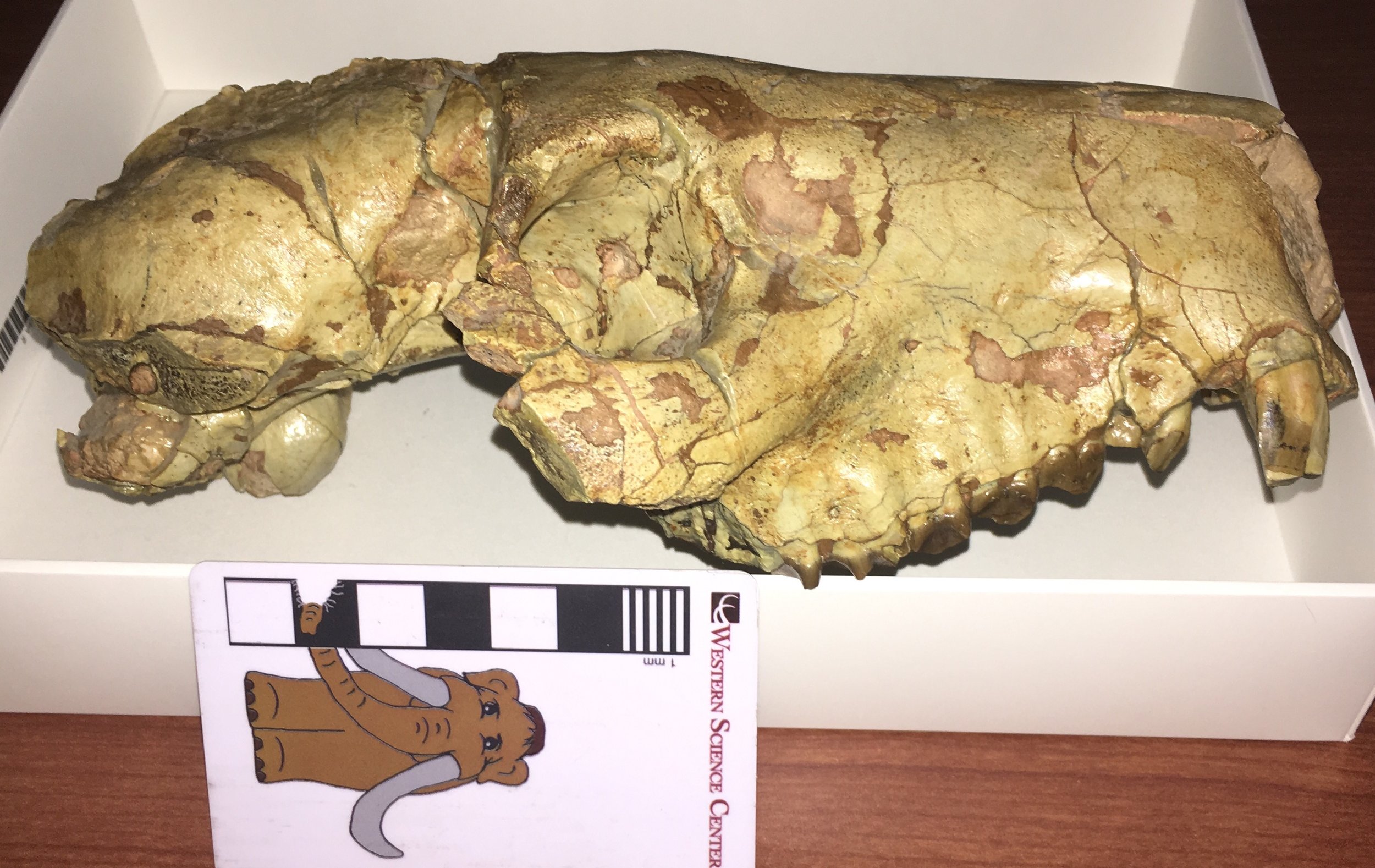 Dorsal view:
 And ventral view:
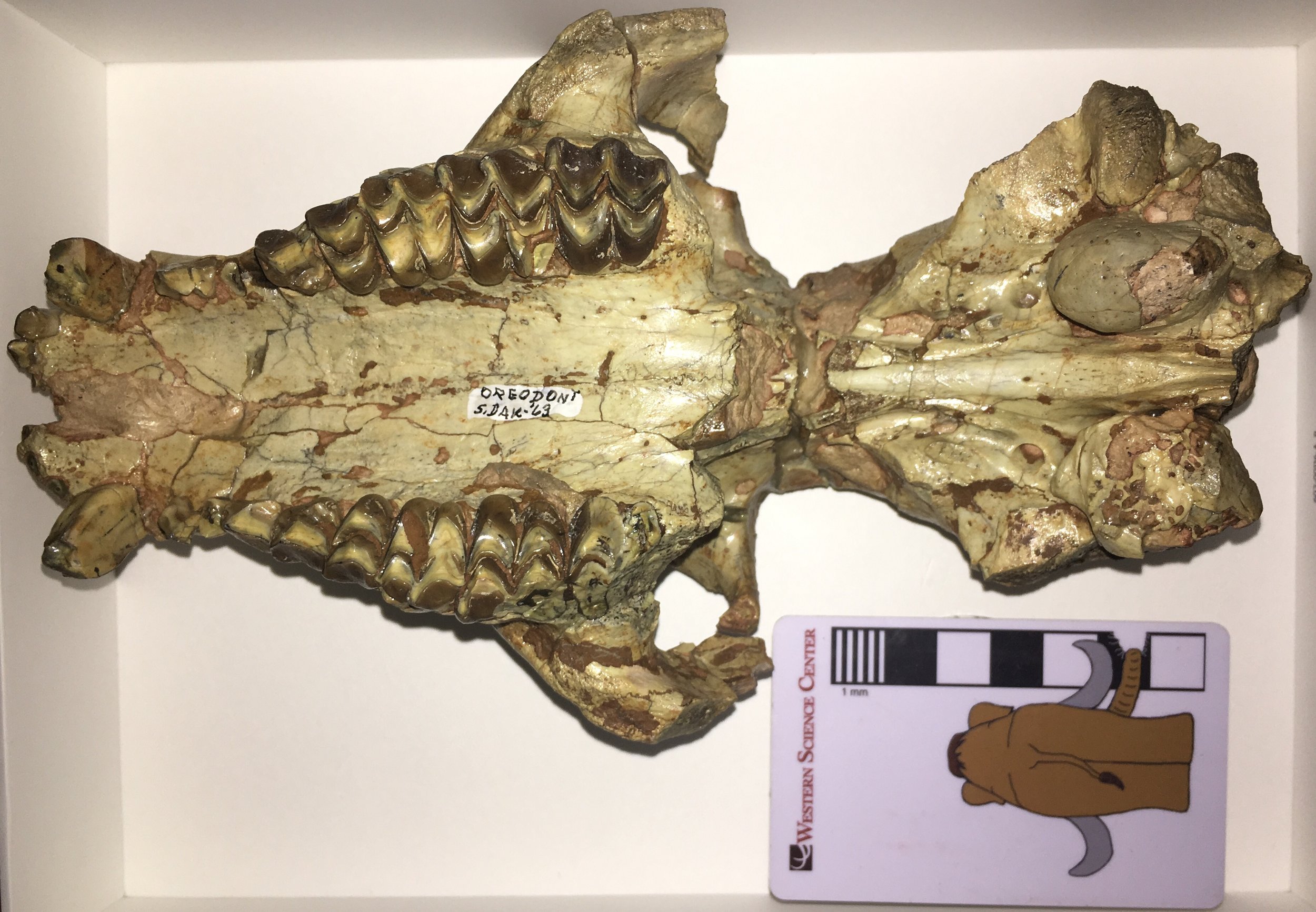 The front of the snout seems to end abruptly, as if part of it has been broken off, but the short, deep skull is actually normal in most oreodonts. If you look carefully at the teeth in ventral view, you can see that the incisors are preserved, confirming that the snout is complete. Based on the size and shape of the skull, I believe this specimen is from Merycoidodon culbertsoni, one of the most common oreodonts in the White River Group. Thousands of specimens have been collected, especially at Badlands National Park and the surrounding areas. The Museum of Geology at the South Dakota School of Mines has many beautiful examples of Merycoidodon on display, including this female that was pregnant with twin calves:
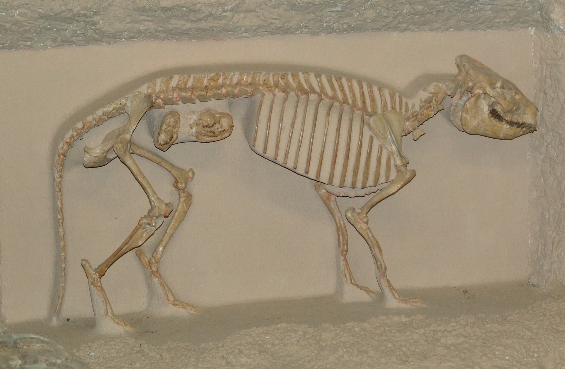 Oreodonts are often described as goat- or sheep-like, which is okay as far as it goes; Merycoidodon was about the side of a sheep. But oreodonts were most common before grasslands became widespread, so they were clearly not grazers in the same sense as sheep. This description also doesn't do justice to the diversity of oreodonts, which included forms that are nearly as large as a cow, as well as some that were probably more like tapirs and hippos in their habits.For more information on oreodonts and the environments they frequented, I encourage you to check out the virtual field trip on the Badlands National Park that Brett and I wrote a few years ago (link at the top right of the page; this requires iBooks on an iOS or MacOS device), or the Badlands National Park Twitter feed (@BadlandsNPS).
Fossil Friday - sloth thoracic vertebra
 Earlier this week, sloth expert Greg McDonald spent several days at the Western Science Center looking at ground sloth material in our collection. I spent that time peering over his shoulder and asking lots of questions. This made the work go more slowly, but it also greatly improved my understanding of ground sloths.At the top is an anterior view of a vertebra from Paramylodon harlani, the most common ground sloth from Diamond Valley Lake. Even though this is a damaged bone, it still shows some important features for understanding sloths.Sloths, along with anteaters and armadillos, are members of the Order Xenarthra, a group that is native to South America. While these animals may seem pretty disparate in their body plans, there are characters that they all share that reveal their relationship to one another. One of the most significant features is reflected in the name of the group; Xenarthra roughly means "alien joint".Most tetrapods have vertebrae that articulate with the vertebrae both ahead of and behind them in the column. Besides the main body of the vertebra (the centrum), there are usually a pair of articulation at the top of the neural canal called the zygapophyses. At the top front edge of the neural arch are the left and right prezygapophyses. The articular surfaces of the prezygapophyses face more-or-less dorsally, and articulate with the postzygapophyses, which are at the top back edge of the neural canal and face ventrally. Below is the dorsal view of a mastodon vertebra featured on Fossil Friday last April, with the prezygapophyses outlined in red (they would normally be symmetrical, but this specimen had been injured):
 Below is an oblique view of the Paramylodon vertebra, looking down from in front (the centrum is partially visible at the bottom of the image):
 Here is the same image, with the prezygapophyses outlined in blue:
 Notice the two articulations outlined in red? Those are the xenarthrous processes (the "alien joints"), which articulate with corresponding points on the posterior edge of the neural canal of the preceding vertebra. The xenarthrous processes are the key feature linking together the various members of the Xenarthra, and are not found in any other group of mammals.Thanks again to Greg McDonald for visiting WSC and giving us so much help with our sloth material.
Fossil Friday - sloth dermal bones
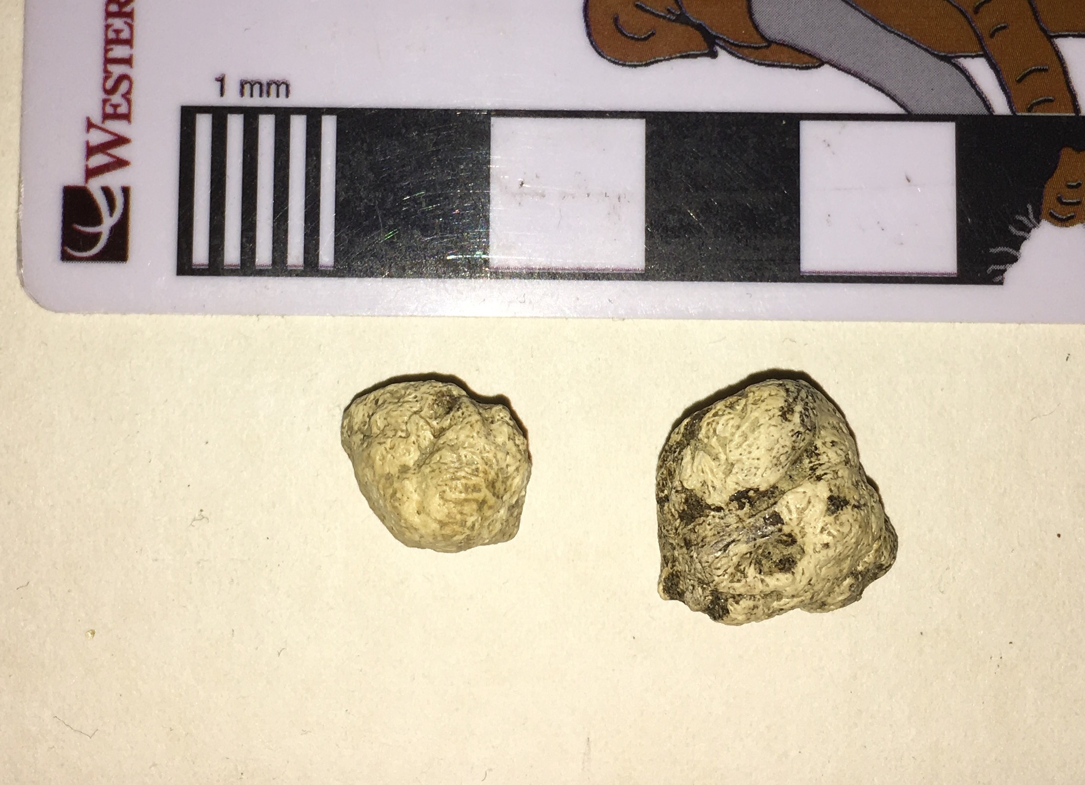 Next Tuesday evening, Greg McDonald is going to give a lecture at Western Science Center on fossil sloths, so for this week's Fossil Friday we have sloth bones!The tiny bones shown here are actually the most numerous sloth element found at Diamond Valley Lake. These are dermal ossicles, bony nodules embedded in the skin of certain species of ground sloths. Below is another view of the same two specimens:
 Of the three sloth species found at Diamond Valley Lake, only one, Paramylodon harlani, is known to have had dermal ossicles. These two ossicles were found associated with a partial skeleton of Paramylodon from the East Dam (part of the jaw of this individual was featured in an earlier Fossil Friday). In fact, the larger ossicle had rolled into one of the empty tooth sockets in the lower jaw.The presence of dermal ossicles in some ground sloths is a bit surprising. The ossicles are generally assumed to provide armor protection, but they are tiny, and it's not clear how extensive they were. While several patches of preserved Paramylodon skin have been found with numerous embedded ossicles, we don't seem to find enough ossicles to cover the entire, or even most, of the body. It's also curious that most ground sloths seem to have gotten along perfectly well without dermal armor, including Megalonyx, which was close to the same size as Paramylodon, overlapped with it in time and space, and was even more widely ranging. Dermal armor is common in some other groups within the Xenarthra (the order that includes sloths), such as the armadillos and glyptodonts. So it's possible that the armor in Paramylodon is a relict, a holdover from some earlier sloth ancestor. But this would mean the earliest sloths should have had armor (as far as I know this is not the case), and that armor was subsequently lost in almost every sloth lineage except for a few species. So for now the origin and function of Paramylodon dermal ossicles remain a bit mysterious.
Fossil Friday - mastodon skull fragment
 After the turmoil of end-of-year administrative duties, I'm now starting to turn my attention back to the Mastodons of Unusual Size Project.The subject of these week's Fossil Friday is a skull fragment that is being added to our dataset.The fragment is shown above in lateral view, with an annotated version below:
 While most of the skull is missing, there is a substantial part of the left front preserved, including the socket for the left tusk, the upper left 2nd molar, and part of the bottom edge of the eye socket. Below is a partial ventral view (part of this side is hidden by the plaster jacket:
 In this view it's clear that the 2nd molar is mostly intact. It's also quite heavily worn; this was a mature mastodon, probably almost as old as Max. It's 3rd molar should have been partially erupted, but that part of the skull is not preserved. The 2nd molar is 121 mm long, which is actually the longest M2 I've measured in a California specimen by a fairly large margin. But it's also only a little over 75 mm wide, making it unusually narrow even for a California specimen. The socket for the tusk is partially crushed, so I'm not confident about measurements of its diameter, but it seems on the small side; it's possible this is a female specimen.
Fossil Friday - Megalonyx tooth
 On January 17, Western Science Center is kicking off a new monthly lecture series. Our first speaker will be H. Greg McDonald from the Bureau of Land Management, who is going to be talking about ground sloths, animals which make up a substantial part of the Diamond Valley Lake fauna.One of the the more rare sloths from Diamond Valley Lake is Megalonyx jeffersonii, which is known from fewer than a dozen bones including the isolated tooth shown above. Sloth teeth are unusual compared to most other mammals. The teeth have no enamel, and instead are composed of different layers of dentine and cementum. There are no deciduous teeth, and the teeth that are present grow continuously through the sloth's life. With no deciduous teeth, no enamel, and very little variation between different tooth position (with the exception of the first tooth and the last tooth in some species), it's actually very difficult to tell which teeth are actually represented in the sloth's jaw.The Megalonyx tooth shown above is a cheek tooth, with the occlusal surface of the tooth at the top. When the tooth first starts growing, it is a simple peg at the the occlusal end. The sculptured surface is entirely the result of wear from food and from occlusion with the opposing tooth, so the usual methods of tracing tooth evolution by examining variation in the occlusal surfaces don't work with sloths.If you're in the area, I encourage you to come by the museum on January 17 for Dr. McDonald's lecture to learn more about sloths.
Fossil Friday - bat jaw
 Earlier this week we had our staff holiday party at the Western Science Center, where we watched The Nightmare Before Christmas. That inspired this week's Fossil Friday topic.Above is the partial right dentary (lower jaw) of a western pipistrelle bat, Parastrellus hesperus. It's shown in lateral view, with anterior to the left, and the black and white bars under it are each 1 mm wide; this is a tiny jaw! The single preserved tooth is the lower third molar. Below is medial view:
 Pipistrelles are not a common component of the Diamond Valley Lake fauna, but their presence is not surprising. They are currently common throughout the southwest, especially in desert areas; we occasionally find them roosting on the museum buildings. While widespread, they are not particularly gregarious, and are usually found alone roosting in rock crevices during the day. Like many other bats, they are voracious predators of insects.Pipistrelles are one of the numerous small animal species present in the DVL fauna that are still common in southern California today. They form an interesting contrast with the large animal fauna, which is almost completely different from what currently lives here.
Fossil Friday - horse skull
 Among the large animals recovered during the Diamond Valley Lake excavation, fully 1/5 of the specimens came from horses; only bison bones were more common among the large animals. There are two species of horse represented at DVL. The smaller species, Equus conversidens, is exceptionally rare in the deposit, and nearly all the recovered specimens belong to the larger Equus occidentalis. Reflecting this, there are a number of E. occidentalis skulls in the WSC collections.The specimen shown above is a partial skull of E. occidentalis seen in ventral view, with anterior to the right. The incisors are missing, but otherwise the right dentition is almost complete. The right teeth are labeled below:
 The only bones visible in this view are the maxillae, which hold the teeth, and a small part of the palatines, which form the curve at the posterior end of the palate (on the left in this image). That curve also forms the front edge of the interior nares, where the nostrils pass through the skull.This individual was an adult horse, with all of its permanent teeth erupted and showing wear. It was recovered from the West Dam area, which includes the most recent Pleistocene deposits in DVL. Carbon dates on samples from the West Dam area were all less than 20,000 years old, and most were less than 14,000 years (Springer et al. 2009).Reference:Springer, K., E. Scott, J. C. Sagebiel, and L. K. Murray, 2009. The Diamond Valley Lake Local Fauna: Late Pleistocene vertebrates from inland Southern California. In L. B. Albright III (ed.), Papers on Geology, Vertebrate Paleontology, and Biostratigraphy in Honor of Michael O. Woodburne. Museum of Northern Arizona Bulletin 65:217-235.
Fossil Friday - whiptail jaw fragment
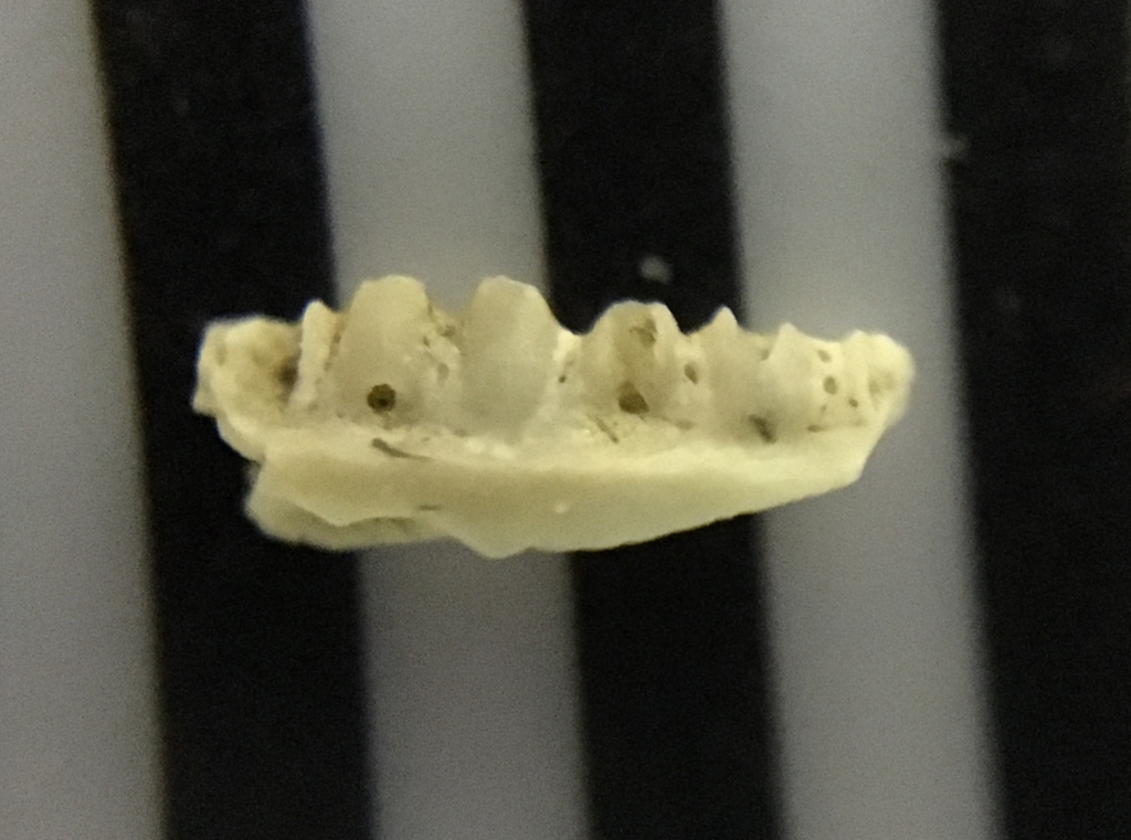 At many sites, screening sediment is among the most effective means of recovering fossils, particularly small bones. Fossils recovered through screening are actually the most common remains from Diamond Valley Lake, and are the source of most of our information about the small animal fauna.The fragment shown above is part of the dentary of a western whiptail lizard, Aspidoscelis tigris (Cnemidophorous tigris in older taxonomic schemes). I'm not sure if this is the left or the right dentary, but it is the medial side, and the bases of four teeth are visible. This is a tiny fragment; the black and white bars are each 1 mm wide.Here's the lateral view:
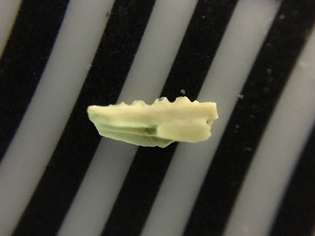 In many lizards the dentary rises higher on the lateral side than the medial side, hiding the bases of the teeth. While four teeth are preserved, the tips of all of them are broken off. This is also apparent in occlusal view (unfortunately not with the best focus; this photo was a bit beyond the limits of my iPhone's camera):
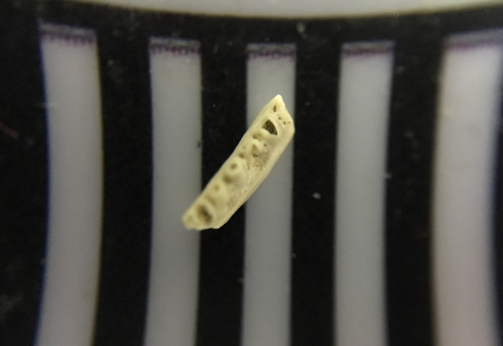 Aspidoscelis tigris is a small lizard with (as its name implies) an exceptionally long tail. The example below was at the Henry Doorly Zoo,hanging out on top of a Gila monster (Heloderma suspects):
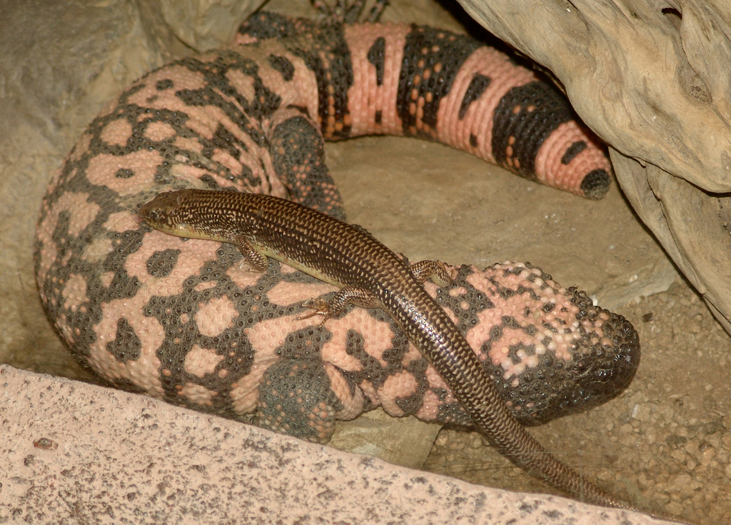 Whiptails are common today in arid and semi-arid parts of the southwest, where they feed on insects and other arthropods such as spiders and scorpions. This specimen was recovered from the East Dam of Diamond Valley Lake.
Fossil Friday - coyote jaw
 The deer bones I talked about a few weeks ago are part of a small assemblage from a housing development in Murrieta, in southwestern Riverside County. Among the other remains in the collection was a partial dentary (lower jaw) from a carnivore.The fragment is a right dentary, shown above in lateral view with anterior to the right. Below is medial view, with anterior to the left:
 And here's the dorsal view, with anterior to the left.
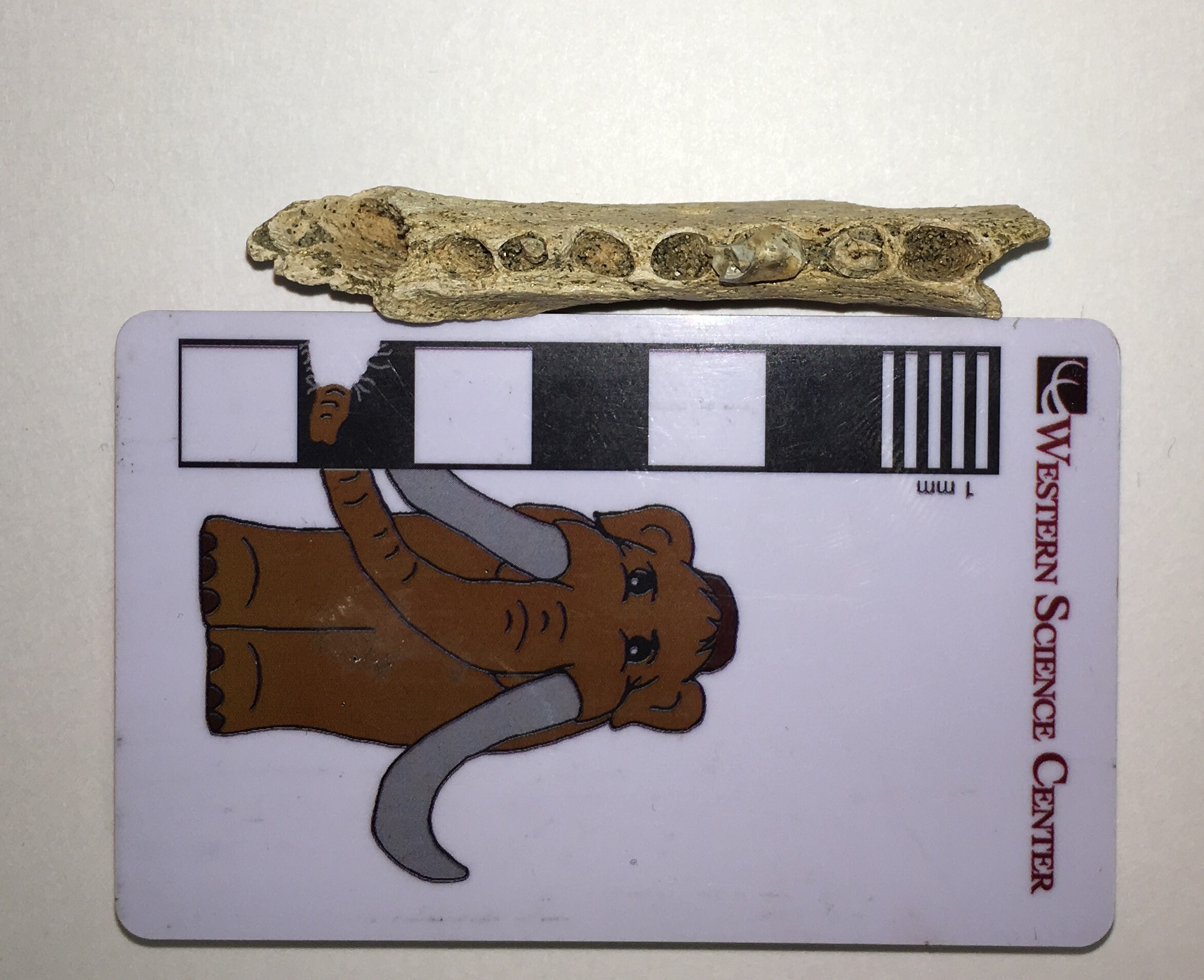 The teeth are mostly missing, but the back half of the third premolar and the anterior root of the fourth premolar are preserved. Below is dorsal view with the sockets labeled:
 This fragment is a good match in both size and morphology for the front half of a coyote, Canis latrans, which have been common in California since the Pleistocene:
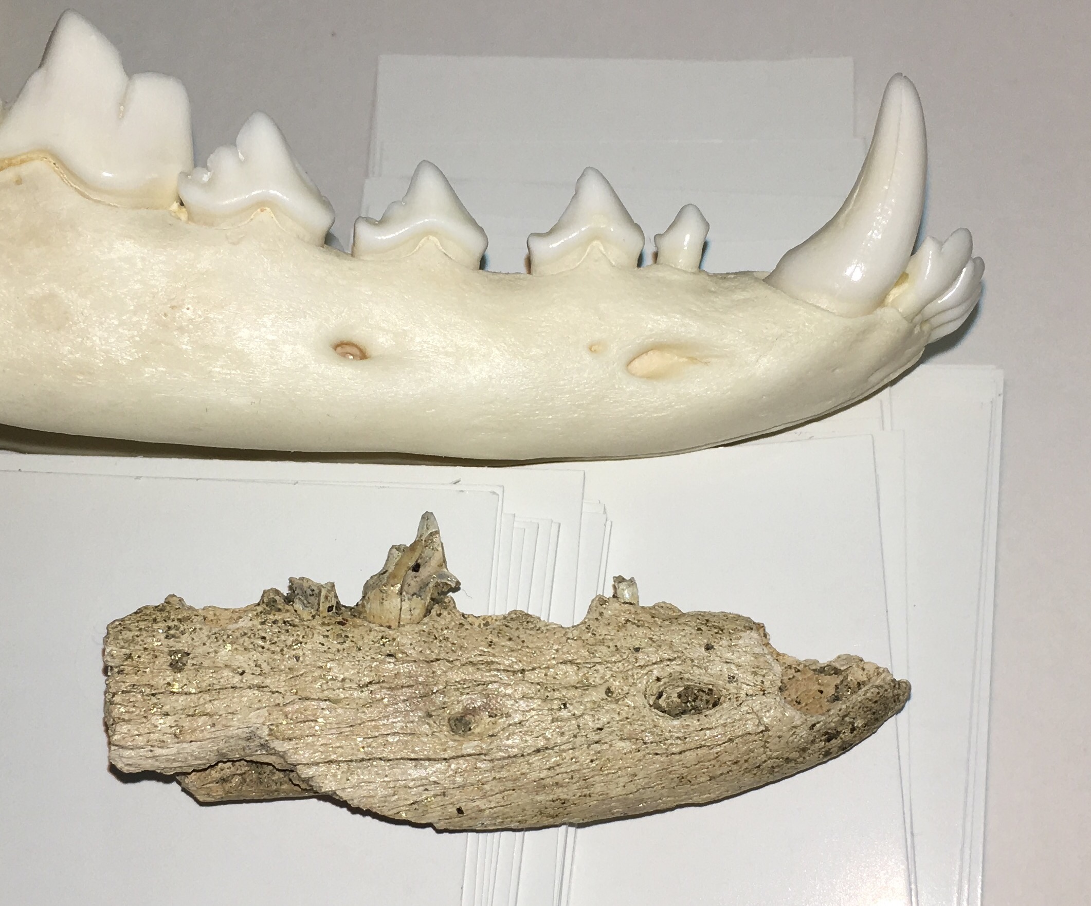

Fossil Friday - Foerstephyllum vacuum colony
 Western Science Center has a relatively small collection of invertebrate fossils, but we've been working over the last two years to increase our holdings of invertebrate specimens. The specimen shown above is a coral colony from the species Foerstephyllum vacuum. It was collected from the Late Ordovician Drakes Formation in Kentucky, from a unit known as the Otter Creek Coral Bed. There are several coral species found in this unit, with Foerstephyllum being the most prominent.Foerstephyllum is a member of a group of corals known as the Tabulata. The tabulate corals were a highly successful and diverse group that were found worldwide during the Paleozoic Era over a span of more than 200 million years. Like so many other groups, the tabulates went completely extinct at the end of the Permian Period 250 million years ago, when roughly 95% of marine invertebrate species went extinct.