

 Almost all the species alive today were also around during the Ice Age. While they may have been filling different niches in the much more diverse Pleistocene fauna, their remains are very recognizable. WSC has a small collection of fossils from Murrieta that include remains from a number of extant species. Among these are two associated forelimb bones from a deer, the humerus (above) and the radius (below, both in anterior view)
 Below are the same two bones in posterior view:
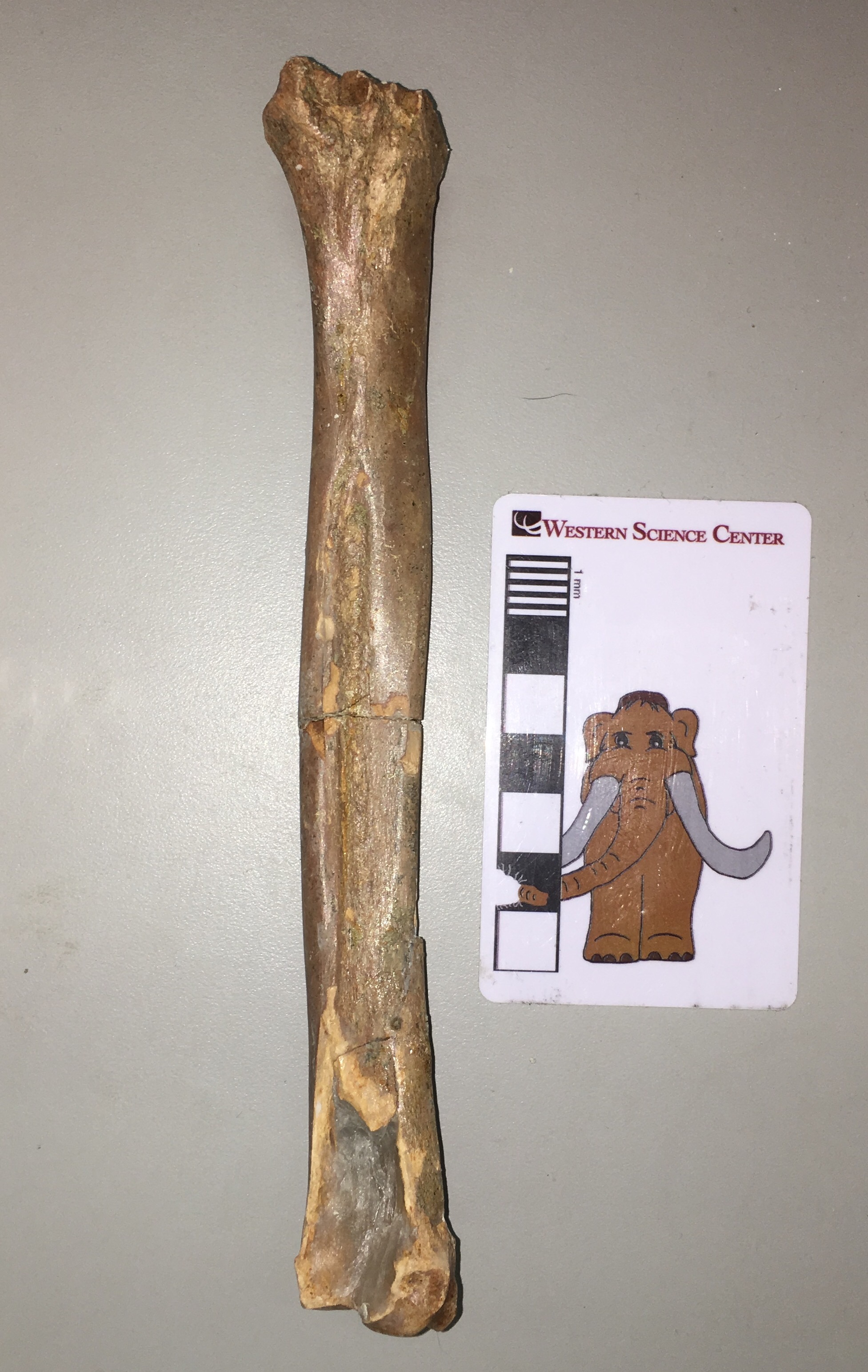 The humerus is missing the proximal 1/3 or so, but is otherwise in good shape. The radius is nearly complete except for a fragment at the distal end. While it's difficult to see in these photos, there is some evidence of gnaw marks from rodents at the distal end of the humerus.Both of these bones are from the left forelimb, and they articulate quite nicely (lateral view, with anterior to the left):
 These bones are pretty much a perfect match for Odocoileus, the genus that includes the white-tailed deer and the mule deer. They are surprisingly small, however, and are a good bit smaller than the female white-tailed deer I was using as a reference. I'm not sure if this has any significance, since it's difficult to draw generalized conclusions from a single specimen.
Fossil Friday - Proboscidean tusk
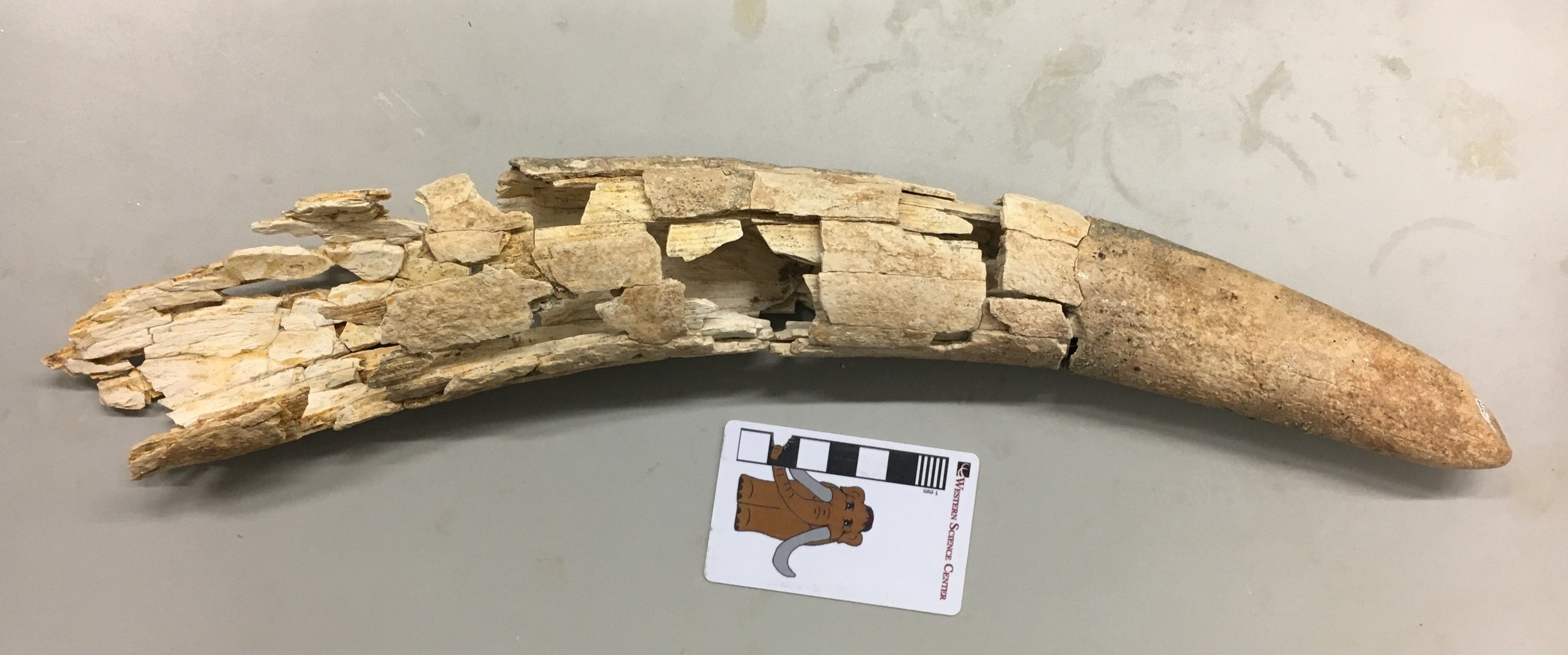 While the Diamond Valley Lake Project lasted for several years, it was still essentially a salvage operation. As a result, many of the larger specimens have only been partially prepared. Our staff and volunteers are gradually working through the backlog.For the last few months our volunteer Nathan has been reconstructing a shattered tusk, consisting of a tray with perhaps 300 fragments. His hard work is paying off, as a pretty nice tusk is beginning to take shape. More than half of recovered fragments have been reattached so far. We're not sure yet if this tusk is from a mammoth or a mastodon, although I'm leaning toward the latter. Assuming it is mastodon, it's either from an immature animal or from a female; adult male mastodons have massive tusks that are much larger than this. The tusk is worn at the tip, and there appears to be more than one wear facet. I'd like to say this indicates an older animal, but ivory is relatively soft and wears rapidly, so even a young animal can have substantial wear at the tip of the tusk.The DVL mastodon collection is dominated by adult males, and we don't have a lot of mammoths at any age, so this tusk represents a relatively rare part of the collection no matter what it ultimately turns out to be.
Fossil Friday - pocket gopher jaw
 This week's Fossil Friday subject is one of the most common Pleistocene vertebrate fossils in the Diamond Valley Lake fauna; the pocket gopher.Pocket gophers are rodents from the Family Geomyidae, which first evolved in North America during the Oligocene or early Miocene. The family diversified in the late Miocene and Pliocene and is now found throughout the New World.Above is the right dentary (lower jaw) from a western pocket gopher of the genus Thomomys. It's shown in lateral view, with anterior to the right. The anterior end is dominated by the huge ever-growing incisor, which is one of the key features shared among all rodents. In medial view (below), the cheek teeth are more clearly visible:
 The back end of the dentary is missing, so the condolences that articulates with the cranium and the coronoid and angular processes where the jaw muscles attach are not preserved. Adult pocket gophers have a total of 5 teeth in each half of the lower jaw, an incisor, the 4th premolar, and 3 molars. In this specimen, the 4th premolar and 3rd molar are missing, which is more obvious in dorsal view:
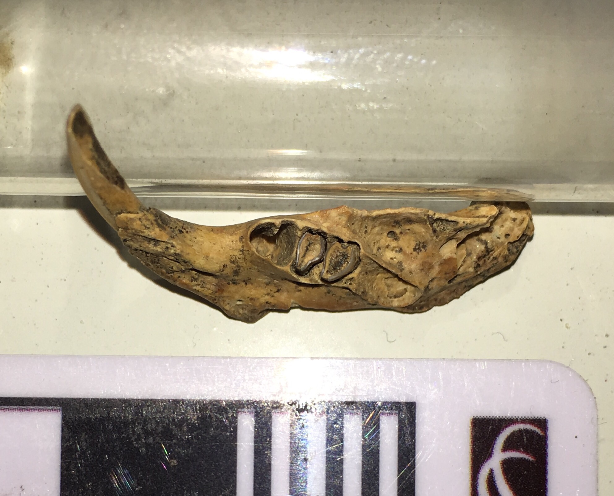 The 4th premolar is a bi-lobed tooth, as is clear from its empty socket. The 1st and 2nd molars are well-preserved and show typical wear for an adult pocket gopher, while the 3rd molar is missing and there's some damage to its socket.Pocket gophers are burrowing animals that make vast tunnel systems where they store their food, but they don't live in large colonies like prairie dogs and other ground squirrels. Thomomys teeth are very common in the Diamond Valley Lake deposits; they are in fact probably the most common vertebrate at DVL, making up nearly 1/3 of all the fossils found there.
Fossil Friday - sloth jaw
 Yesterday was International Sloth Day (with sloth in this case being a noun, not an adjective)! That's a nice day to celebrate at Western Science Center, because Diamond Valley Lake is the only locality in California with three different species of ground sloths. By far the most common sloth at DVL is Harlan's ground sloth, Paramylodon harlani. Shown above is a Paramylodon partial lower jaw. This is the anterior end of the right dentary seen in dorsal view, with anterior to the left. The long straight edge at the lower left is the mandibular symphysis, where the right and left dentaries attach to each other (essentially the chin). The bases of two teeth are visible at the upper right.Here's a lateral view:
 This time the anterior end is on the right. In this view it's clear that the teeth are broken off, and barely protrude above the bone. This is presumably breakage that occurred after the animal died, since the breaks are jagged.This fragment makes an interesting comparison with one of the other DVL sloths, Megalonyx jeffersonii. Paramylodon seems to have a longer, more slender jaw than Megalonyx and lacks the latter's enlarged, chisel-like incisors. That's not entirely surprising; while Paramylodon and Megalonyx are both sloths, they are only distantly related to each other.
Fossil Friday - horse jaw
 While the majority of Ice Age fossils in Riverside County are from the Diamond Valley Lake excavation, there are a few other productive Pleistocene sites. As with DVL, these have mostly been found during construction projects. The specimen shown above was recovered from The Promenade shopping mall in Temecula.While badly crushed, the specimen is the tip of the lower jaw of a horse. It's shown in dorsal view, with anterior to the left. Mammal lower jaws are made up of two bones, the left and right dentaries. In most species the dentaries are fused together at the chin, in what's technically called the mandibular symphysis; that's what's preserved here.There are actually portions of 8 teeth present in this fragment, although they're poorly preserved. In the marked-up image below, the red areas are indicating the positions of the 6 lower incisors (3 on each side), while the blue arrows are pointing at the bases of the canines (
"wolf teeth" in horse terminology Eric Scott pointed out to me that the "wolf teeth" are actually vestigial 1st premolars, not the canines, so that while these teeth are canines they are not wolf teeth):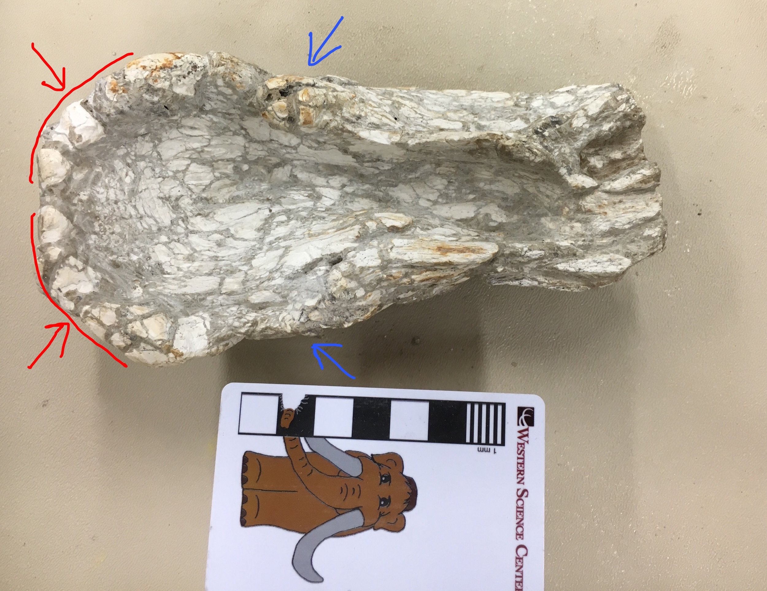 WSC volunteer Joe has been cleaning this specimen, and is working on other associated fragments. It looks like we probably have additional material from this jaw, which I'll post once it's further prepared.
The role of museums
 Inspired by Brian Switek’s recent article in Aeon, I was reminded of a post I wrote several years ago for “Updates from the Paleontology Lab” about different types of institutions that describe themselves with the term “museum”. What follows is an updated and edited version of that post.Over the course of my career I’ve had the opportunity to visit numerous natural history museums for a variety of reasons, as a researcher, a public lecturer, a tourist, and, of course, as an employee. The institutions are highly diverse in terms of their missions, and it’s instructive to reflect on those differences. The points I’m going to make are largely anecdotal, but should serve to give a general idea of the different types of natural history museums.During my time in Virginia, I made several visits to the Science Museum of Virginia (SMV), located in Richmond. SMV has a very large exhibit area and I was always impressed with the range and quality of hands-on interactives in their exhibits, but there were very few specimens on display.
 Extinction exhibit at the Science Museum of Virginia.SMV is an excellent example of a science education center. These institutions generally have few (if any) collections, and usually what collections they have are used in education rather than research. They are generally staffed by educators, with few if any active research scientists. The primary audiences are families with school-age children or schools themselves, especially elementary and middle schools, and this is reflected in the types of exhibits that are selected. This is not to say that adults can’t benefit or enjoy these exhibits - just that they are not the primary audience.A number of colleges have campus museums; two that I’ve visited recently are the Joseph Moore Museum at Earlham College and the Museum of Earth Science at Radford University. While it used to be fairly common for small colleges to maintain museums, they fell into disfavor in recent decades; fortunately they may be experiencing a bit of a renaissance.
 Allosaurus cast on display at the Joseph Moore Museum.Campus museums are almost as eclectic as the colleges that run them, but they do tend to have a few things in common. Collections at these museums are primarily for teaching (although there are exceptions; we’re including several Joseph Moore Museum specimens in the “Mastodons of Unusual Size” project). Most of these museum have few or no paid full-time staff, and often are run part-time by a faculty member. Most of the museums’ operational activities, including their programs, are conducted by students. Often these institutions serve as teaching museums, and are where many career museum professionals got their initial exposure to museum operations.
 Collecting mastodon data at the Joseph Moore Museum.Finally, there are research museums such as the Western Science Center. Many research museums are operated by federal, state, or local governments (I previously worked at the Virginia Museum of Natural History, a state-funded research museum). Some, such as the Florida Museum of Natural History, are departments within research universities, which are also often state government-supported. A few, such as the American Museum of Natural History and Western Science Center, are stand-alone non-profit institutions operated in most cases by private foundations. While research museums may have different origins and associations, they all share a common feature: a permanent collection of research specimens. The primary function of these museums is to preserve the specimens and associated data that form the basis of natural science. These museums will usually have a written collections policy that defines the scope of the collection, how specimens are acquired, and how they are made available for study. The collections supersede any particular staff member; the curators and collections staff are there primarily there to support and maintain the collections, not the other way around.
 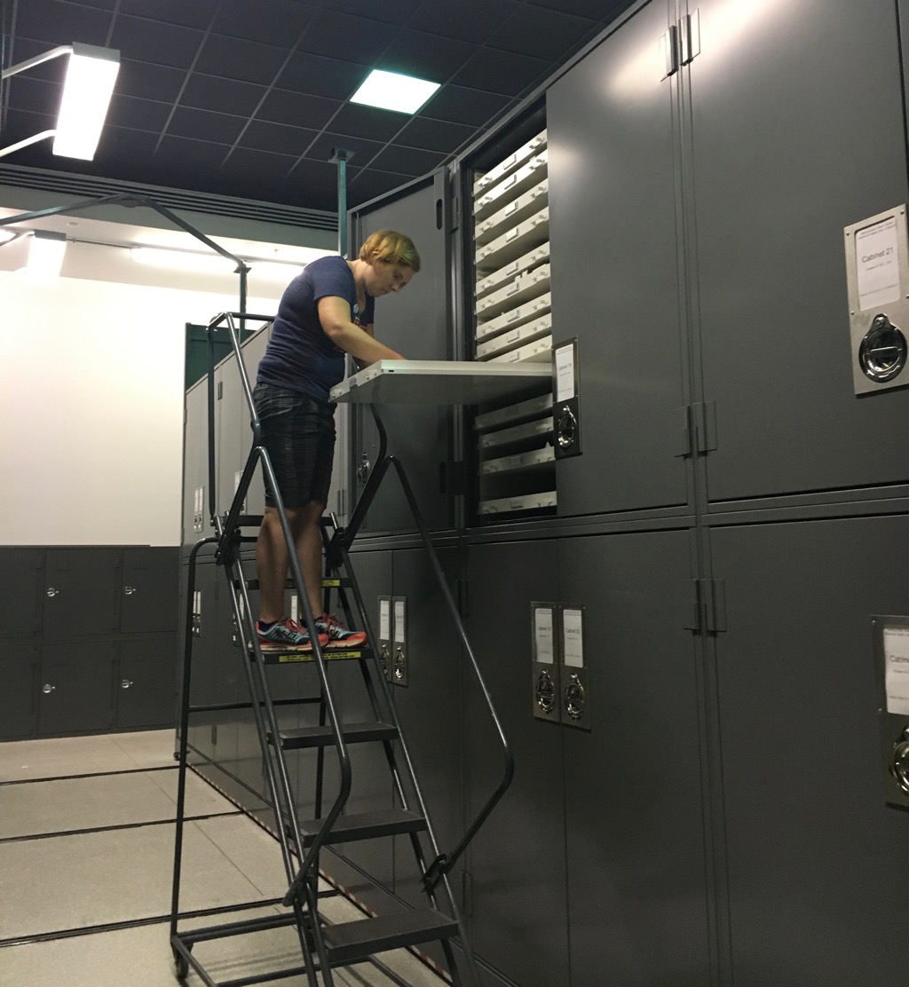 Dr. Katy Smith working in the collections repository at the Western Science Center.Specimens in a research collection are selected for their scientific value. Many of these specimens may be visually appealing, but that’s not the reason they’re acquired. In fact, the majority of a research museum’s collections are rarely if ever viewed by anyone except specialists who incorporate the data from those specimens into their research. In this respect, research museums stand apart from science education centers and teaching museums.Of course, these are broad categories that tend to merge into one another. Many museums have a close relationship with colleges, even if they aren't actually operated by a college. Some science education centers have small but significant research collections (the Boonshoft Museum of Discovery is a science education center where we collected mastodon data). Nearly all museums put a lot of effort into public education, regardless of their primary focus. Nevertheless, I'd suggest that most natural history museums tend to fall closest to one of these three categories.With such a range of missions and holdings, why are all these disparate institutions called museums? There is one feature that they all tend to share: exhibits. Even though the primary function of a research museum is maintaining collections, exhibits are a critical supporting part of that mission. To extend an analogy that a friend once suggested to me, why does a football stadium or a theatre have seats? Why do we have art galleries? All the players, actors, or artists require is a field, stage, or easel. The seats in a stadium or theatre are for everyone else – those who aren’t athletes or actors. The theatre, art gallery, and playing field are venues for non-specialists to experience these activities. Likewise, museums are the stages that scientists use to present their work to others. The vast majority of scientific research is funded, in one way or another, by the public. We have an obligation to make the fruits of our scientific endeavors available to as many people as we can, and particularly to those who pay for them.With exhibits serving as one of the significant public faces of research, it’s easy for the public to become misled about the function of museums and exhibits, particularly when the term “museum” is used for a variety of institutions. It can seem to a visitor that the only reason museums exist is to have exhibits. In the case of a science education center such as SMV, this is actually the case; they are an institution that exists to provide science education, primarily through exhibits. But the knowledge that’s imparted in SMV’s exhibits came from somewhere; someone had to collect and interpret the raw data that led to those discoveries. That was done in university and government research facilities…and in research museums. Institutions such as WSC don’t just provide the scientific knowledge described in our own exhibits, but also for the exhibits in other venues that don’t perform research or maintain collections. For research museums, we don’t acquire collections in order to supply the exhibits; rather, the exhibits exist to teach the public about the significance of our collections. All these different types of museums may share exhibits as a common point, but they perform different, yet complimentary, roles in the advancement of science.
Fossil Friday - Max's pelvis revisited
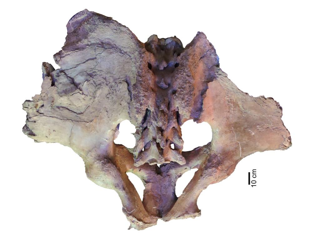 Western Science Center's largest mastodon, Max (@MaxMastodon on Twitter) has been getting a lot of attention over the last year. Besides getting CT scans and figuring prominently in the "Mastodons of Unusual Size" project, this October marks 21 years since Max was discovered. Max is also one of the WSC's major exhibits, and at our recent Science Under the Stars fundraiser we successfully raised funds to add new content to Max's (and other) displays, discussing some of the new things we've learned.Max is not only the largest mastodon known from California, but is also pretty large when compared to mastodons from other parts of the country, so we've long assumed he was male (male mastodons tend to be larger than females). But there are several parts of the skeleton where we can take direct measurements to have greater confidence when identifying Max's sex. One of these areas is the pelvis, which as I've discussed in a prior post is one of the well-preserved parts of Max.Lister (1996) showed that in mammoths males and females consistently differ in the ratio between two pelvic measurements, the pelvic aperture width and the minimum width of the ilial shaft, as shown in the image below:
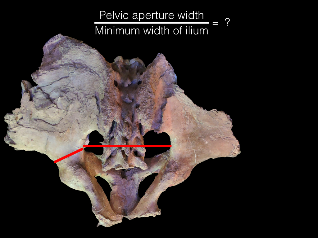 In female mammoths, this ratio is greater than about 2.6, while it is less than 2.6 in males. Hodgson et al. (2008) and Fisher (2008) found that a similar relationship holds true for mastodons. In Max this ratio is 2.23, which is well on the male end of the spectrum. This is, of course, what we expected to find, but it's still necessary to go through the steps and confirm the results, rather than just assuming we'll get the expected answer.One of the new interactive stations funded by our Science Under the Stars fundraiser is going to feature the methods we used to determine Max's sex, including pelvic dimensions. Below is a draft video explanation of the pelvic measurements we put together for the fundraiser, narrated by WSC volunteer Catalina Lauridsen:[youtube https://www.youtube.com/watch?v=Zu4rOYtp9Ng]Tomorrow at WSC we're hosting the first Ice Age Soirée, and over-21 party celebrating 21 years since Max's discovery. Tickets are $40 and include dinner and drinks, as well as admission to the museum.References:Fisher, D. C., 2008. Taphonomy and paleobiology of the Hyde Park mastodon. In Allmon, W. D. and Nester, P. L., eds. Mastodon Paleobiology, Taphonomy, and Paleoenvironment in the Late Pleistocene of New York State: Studies on the Hyde Park, Chemung, and North Java Sites. Paleontographica Americana 61:197-289.Hodgson, J. A., Allmon, W. D., Nester, P. L., Sherpa, J. M., and Chimney, J. J., 2008. Comparative osteology of Late Pleistocene mammoth and mastodon remains from the Watkins Glen Site, Chemung County, New York. In Allmon, W. D. and Nester, P. L., eds. Mastodon Paleobiology, Taphonomy, and Paleoenvironment in the Late Pleistocene of New York State: Studies on the Hyde Park, Chemung, and North Java Sites. Paleontographica Americana 61:301-367.Lister, A. M., 1996. Sexual dimorphism in the mammoth pelvis: an aid to gender determination. In Shoshani, J. and Tassy, P. eds. The Proboscidea: Evolution and Paleoecology of Elephants and their Relatives. Oxford University Press, pp.254-259.
Fossil Friday - vole tooth
 The rodent family Cricetidae is a diverse group that includes animals such as muskrats, pack rats, and hamsters. That diversity is reflected in the Diamond Valley Lake deposits, which contain several different cricetids including one of the most common members of the family, the voles.Voles are small rodents that look vaguely similar to house mice (which are an invasive species in North America and belong to an entirely different family). Below is an example of a meadow vole, Microtus pennsylvanicus, which is found in northern and eastern North America (this one is from Virginia; the small white objects are blowfly eggs):
 As with so many mammals, the most commonly preserved fossil remains of voles are their teeth. At the top of the page is a right lower third molar in labial view; the entire tooth is about 3 mm tall. Below is the lingual view of the same tooth:
 The series of ridges on the side of the tooth are expressed on the occlusal surface as complex loops of enamel. Below is the occlusal view (sorry for the focus, I'm experimenting with a new macro lens for my iPhone):
 This pattern of alternating ridges of enamel with areas of dentin serves to keep the tooth sharp as it wears down, and versions of this have evolved independently in almost every group of mammalian herbivores, from rodents to elephants. It's especially common in groups that feed on highly abrasive plants such as grasses, which are in fact the preferred food of voles.The DVL tooth probably comes from Microtus californicus, a species that it still widespread in California except in very arid regions. While probably not as common as the fellow cricetid Neotoma, voles are still a significant component of the DVL fauna.
Fossil Friday - bison jaw fragments
 Some of the species in the Diamond Valley Lake deposits are common enough that it's actually possible to get some idea of intraspecies variability, including growth-based (ontogenetic) differences. Bison are not as common at DVL as horses, but there are still enough specimens to look a bit at age profiles. The specimen above is the anterior end of the lower jaw, with portions of both dentaries. This is a dorsal view, with anterior to the left. Below is a lateral view of the left dentary:
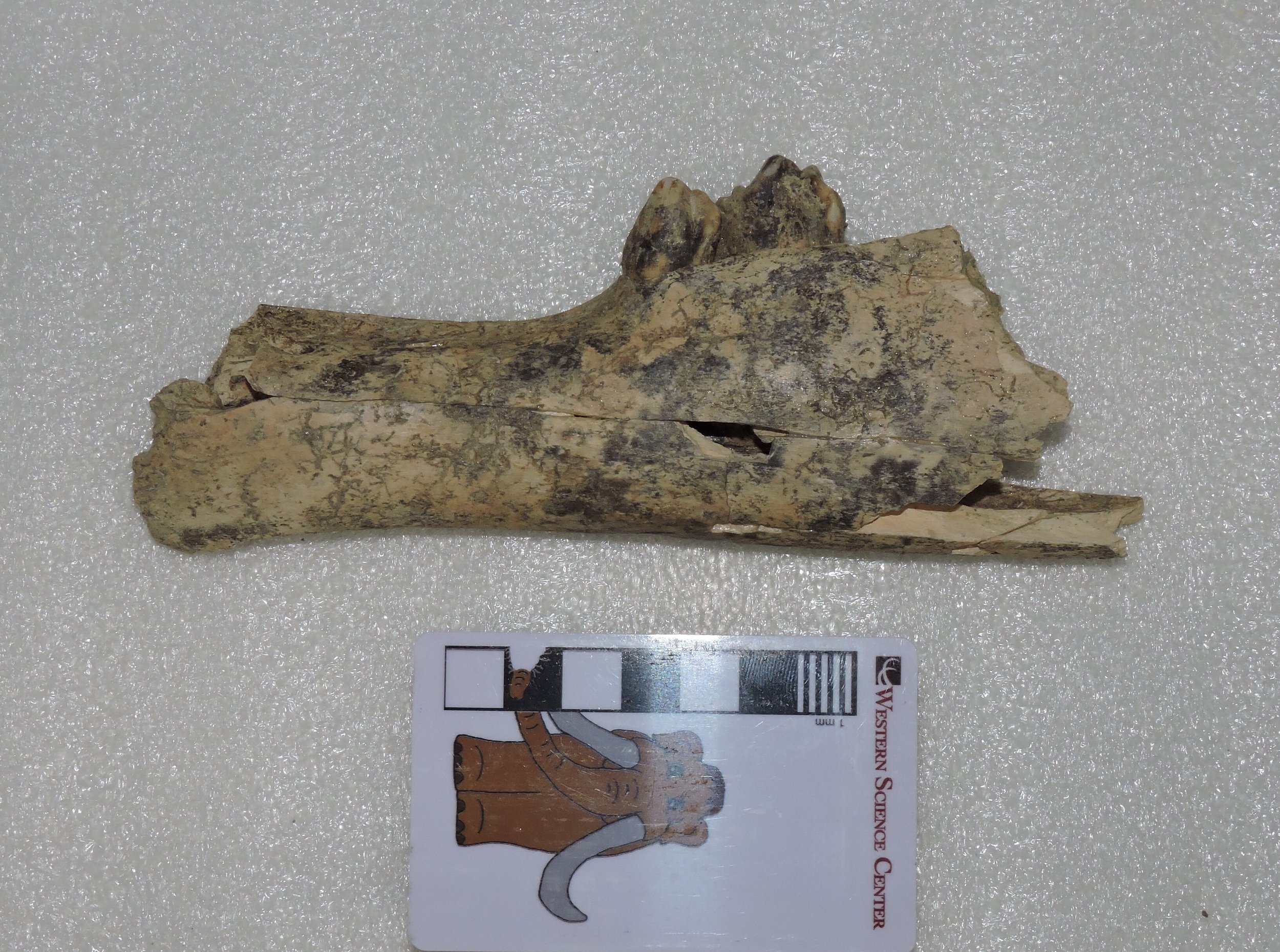 ...and here's right dentary in lateral view:
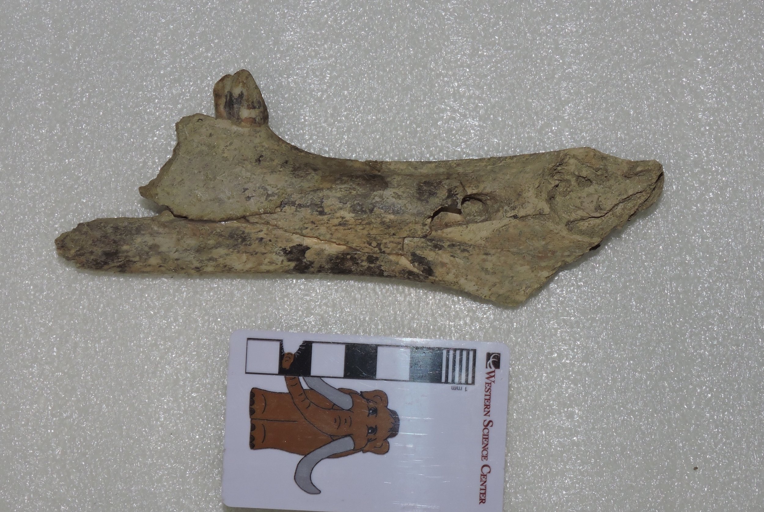 Three teeth are preserved, the right 2nd premolar and the left 2nd and 3rd premolars, none of which have any strong indications of wear. The jaw sections are also quite small compared to other bison in our collection. That raises a question: are these teeth permanent premolars, indicating an animal that was maybe 3 or 4 years old, or are they deciduous premolars, in which case it was probably only a few months old? The small size of the jaw actually makes me suspect the latter. I hope at some point to get x-ray images to see if the permanent premolars are present inside the jaw, although this animal could be so young that they hadn't started to develop yet.
Fossil Friday - Carnivore traces
 In any large collection of vertebrate fossils, one of the more common specimen labels will be "unidentified bone fragment". But even an unidentified fragment can provide useful information. The bone shown above, found near the East Dam of DVL, has the following, rather uninspiring label: "Mammalia (larger size), unidentified bone fragment". This might be part of a transverse process or neural spine from a vertebra, and if so is could be from anything from a horse to a juvenile proboscidean. But there are also other possibilities; about the only things we can rule out are animals like rodents and rabbits that don't have any bones at all that are this large.So why is this bone interesting? Note the grooves visible in several places along the margin. Here are some views from different angles:
 These nearly-parallel grooves are consistent with gnaw marks from a carnivoran, so this fragment is covered with trace fossils. We can't say with certainty what type of carnivoran made the marks, but they are not particularly large, so it's unlikely we're looking at a huge animal like Arctodus. A medium-sized carnivoran such as a black bear, dire wolf, or coyote is a more likely possibility, or perhaps even a badger; all of these are known from the Diamond Valley Lake deposits.As I've mentioned in previous posts, carnivoran bones are quite rare in the DVL deposits. But we do see evidence of their activity. We haven't yet catalogued all the bite marks that are present on DVL bones, but I suspect we actually have more carnivoran trace fossils than bones in our collection.
Fossil Friday - seven bone fragments that built a museum
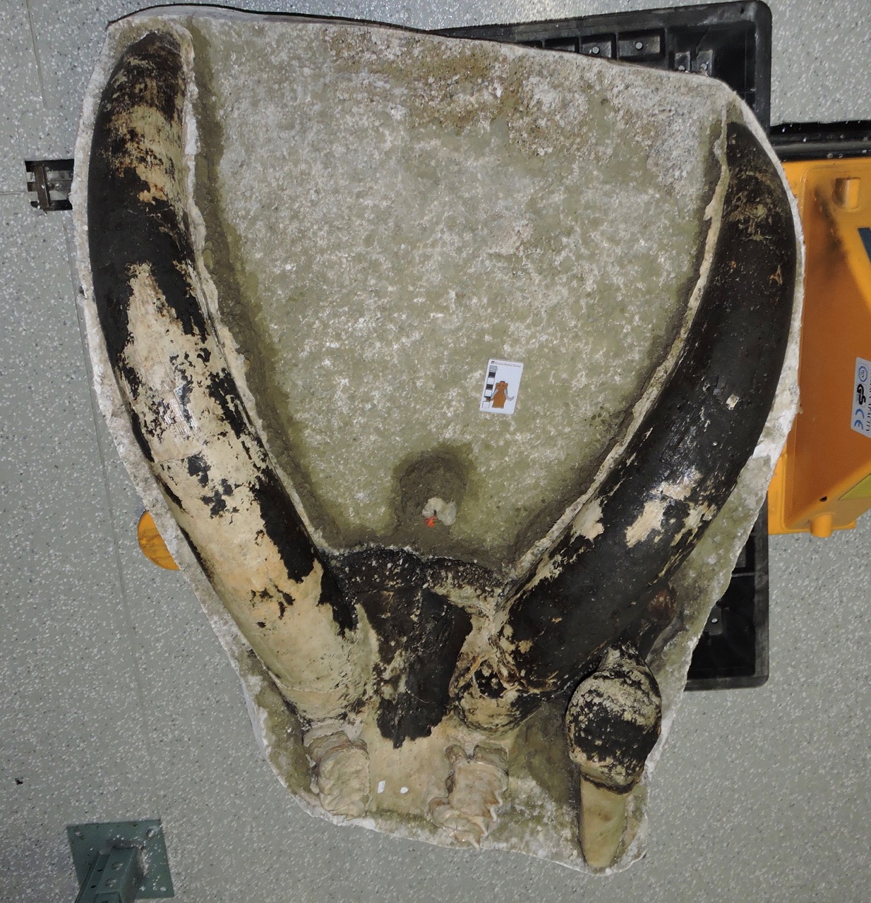
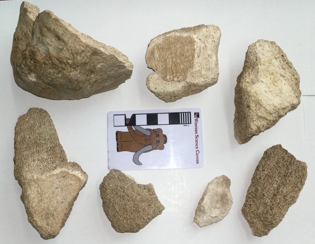 Tomorrow night the Western Science Center is holding the annual Science Under the Stars fundraiser. This year is a particularly special event, because the museum is celebrating its 10th anniversary; we opened to the public for the first time on October 15, 2006.WSC has grown over the last decade into a major regional museum and has brought in collections from a variety of locations, but the original reason we were founded was to house the collections recovered during the construction of Diamond Valley Lake and its related projects, such as the San Diego Canal and Domenigoni Parkway. These specimens still make up the majority of our collections.The seven bone fragments shown above we discovered by archaeologist Melinda Horne on 7 June 1993 along the path of the San Diego Canal. They don't look like much, but one thing is immediately obvious; they're pretty big fragments. The only animals found in California today (outside of zoos) that are even remotely close to being large enough to produce bones like these are horses and cows , and these fragments seem too large even for those. The largest fragment is a bit better preserved than the others; here's a different view:
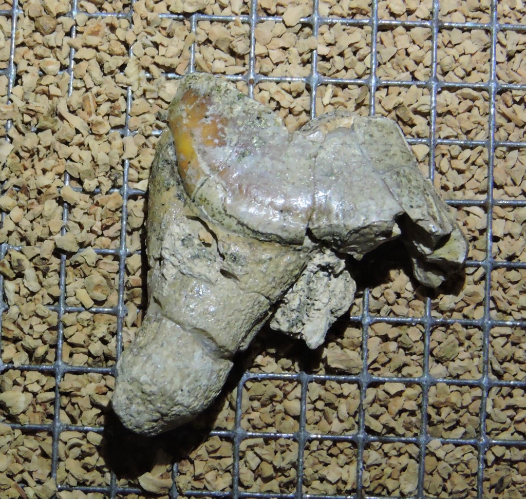
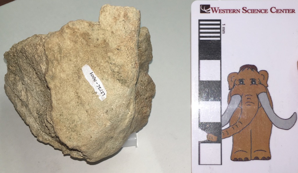 The large, flat surface (where the number is painted) is an articular surface, where this bone articulated with another bone. Even though it's incomplete, there's enough preserved to identify this as the joint surface on the shoulder blade (technically, the glenoid fossa of the scapula). And that gives us another clue; this bone is far too large to be the glenoid fossa from a horse or a cow. The only common things in California this big are extinct Pleistocene elephants. This bone established the presence of Ice Age deposits in the area.This was noted by R. L. Reynolds from the George C. Page Museum, who tentatively identified the specimen as a mammoth, as written on an index card stored with the specimen:
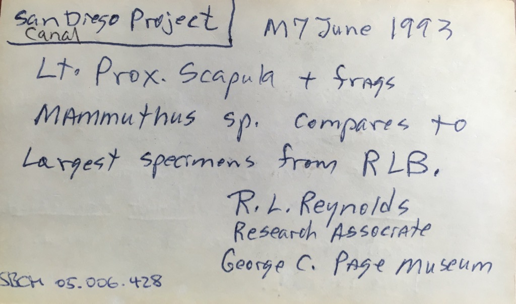 Several years later, Eric Scott (who is now at the Cooper Center, and is one of my collaborators on the Mastodons of Unusual Size project), examined the specimen and considered it more likely to be from a mastodon than a mammoth (his notes were written on the back of the same index card):
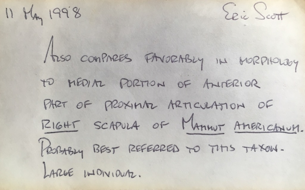 To give an idea of what Eric was seeing, here's a glenoid fossa view of the Watkins Glen mastodon from New York (this image is modified from Hodgson et al., 2008)...
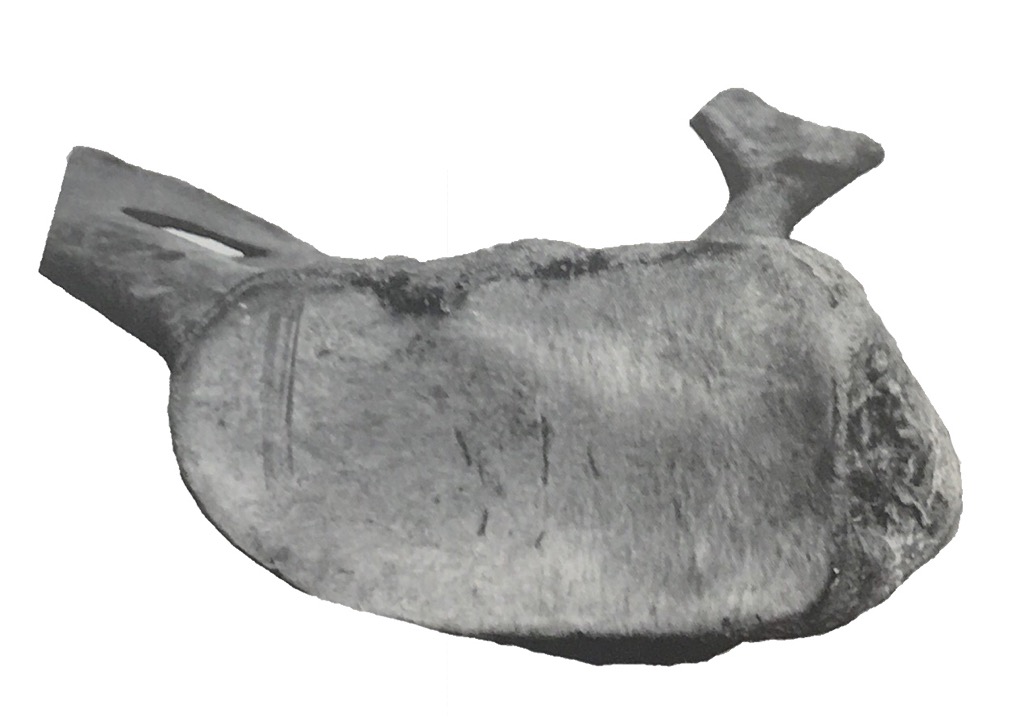 ...and here's the same image with the California fragment scaled to the same size and superimposed:
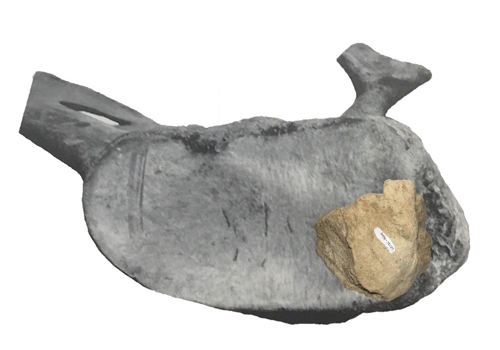 That's a pretty good match, and I agree with Eric that this is probably from a mastodon, the first from the project.These seven fragments put paleontologists and engineers on alert that there could be additional Pleistocene fossils uncovered during the construction of the reservoir. As the project progressed, more specimens were uncovered, and 2 years after the initial find Max was discovered. Over the next decade more than 100,000 fossils were recovered in the area, a collection so large that a new museum was built to house it, and tomorrow night we'll celebrate that museum's tenth anniversary.A fossil doesn't have to look impressive to be important. Reference:Hodgson, J. A., W. D. Allmon, P. L. Nester, J. M. Sherpa, and J. J. Chimney, 2008. Comparative osteology of Late Pleistocene mammoth and mastodon remains from the Watkins Glen Site, Chemung County, New York. In Allmon, W. D. and P. L. Nester (eds.), 2008. Mastodon Paleobiology, Taphonomy, and Paleoenvironment in the Late Pleistocene of New York State: Studies on the Hyde Park, Chemung, and North Java Sites. Palaeontographica Americana 61:301-367. Available for purchase.
Fossil Friday - Xena the Mammoth's femur
 Mastodons were the most common proboscideans in Diamond Valley, but they weren't the only ones there. Columbian mammoths (Mammuthus columbi) were also present, although in much smaller numbers. The most complete Columbian mammoth from DVL is Xena, whose partial skeleton is on permanent exhibit at the Western Science Center.Xena's remains include the left femur (thigh bone), shown above in anterior view with the proximal end on the left. As is typical of Columbian mammoths, the femur is relatively long and slender when compared to mastodons. While adult mammoths and mastodons were probably pretty comparable in terms of weight, mammoths were quite a bit taller because of their long legs, which were proportioned more like those of modern elephants than the relatively short, thick legs of mastodons. Xena was not particularly tall for a Columbian mammoth, but was at least as tall as Max, the biggest mastodon known from California.
 While Xena's femur is well preserved, it is missing both the proximal and distal ends.These were lost because they had not yet fused to the rest of the femur, indicating that Xena was still growing (even at 3 m tall!). This is also borne out by her teeth; Xena's 1st molars were in wear, but the 2nd molars were completely unerupted, suggesting that she was probably only about 18-20 years old.
Fossil Friday - dire wolf tooth
 Today is National Dog Day, and while the day is primarily honoring domestic dogs (Canis familiaris, or Canis lupus familiars), it seems fitting to also recognize their wild ancestors and cousins.The Diamond Valley Lake deposits only produced a handful of carnivores bones, but several canids are included among their numbers. There are a few individual bones and teeth from dire wolves (Canis dirus), the same species that is so spectacularly common at Rancho La Brea, 80 miles to the west.The partial tooth shown above is currently on exhibit at the Western Science Center, so rather than open the exhibit case (a 3-hour exercise) I instead photographed it through the glass. This rather large tooth is a carnassial, shearing teeth developed to varying degrees among all the carnivorans. The upper carnassials are modified 4th premolars, while the lower ones are the first molars. These teeth occlude like a pair of scissors, and are effective at chopping up flesh from prey animals.While it's difficult to do a detailed examination through glass, I'm pretty sure this is the upper right 4th premolar, seen in lingual view, with the anterior edge broken off and the point worn away. Compare it to a modern coyote upper P4 in the same orientation, below:
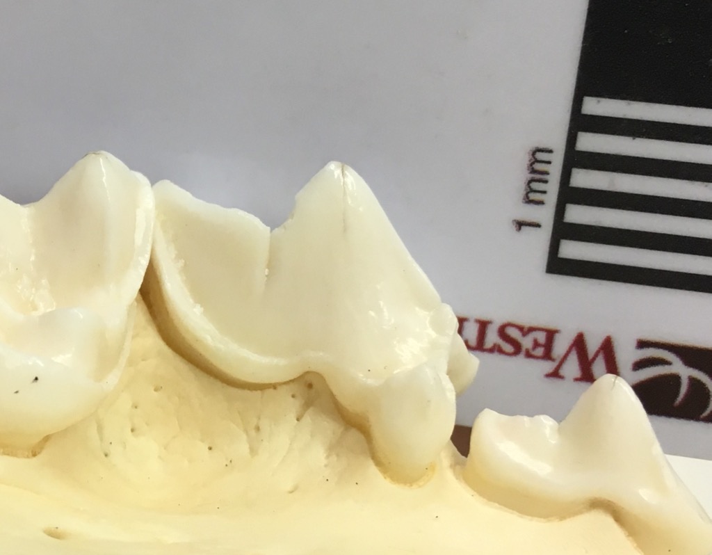 So if you're celebrating National Dog Day, maybe take some time to visit your local natural history museum and remember our canine friends' extinct relatives!
For those in Southern California, the Western Science Center will be attending NerdCon at the California Center for the Arts in Escondido this weekend. There will be opportunities to take selfies with Max, and we'll have numerous replicas on display (including a dire wolf skull). You'll also be able to purchase detailed replicas of some of our fossils, so if you're attending NerdCon make sure to stop by our booth!
Fossil Friday - Pupilla muscorum
 To human eyes, the most noticeable parts of every ecosystem are the big, charismatic organisms; there's a reason the blog is called "Valley of the Mastodon". But in terms of numbers of individuals and, usually, total biomass, small organisms actually dominate ecosystems. That's often reflected in the fossil record as well.Pupilla muscorum is a tiny, air-breathing terrestrial snail (snails are one of a fairly small number of organisms to successfully invade the land). The black bars under the shell shown above are each 1 mm wide, so the entire shell is less than 4 mm long. Shells from this species are quite common in the Diamond Valley Lake deposits; below are the specimens recovered from a single DVL locality:
 Today, P. muscorum is found across the northern hemisphere, but especially in Europe. It appears that many of the modern North American occurrences are actually descended from recent invasive European populations, and there is some debate as to whether or not native North American specimens such as the ones from DVL are actually the same species. The shells are extremely similar, so genetic testing of modern populations will probably be necessary to answer this question.
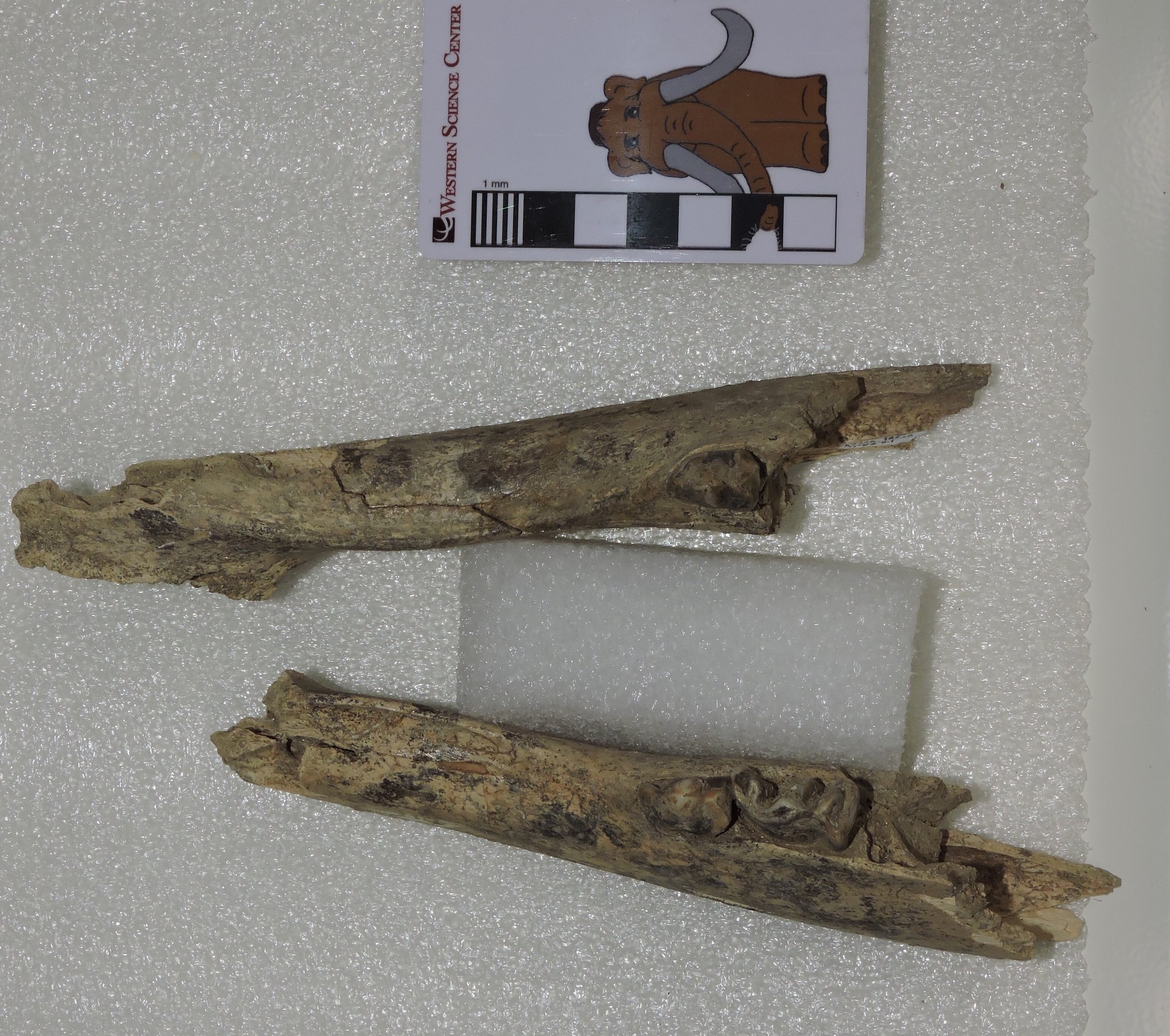
Fossil Friday - mastodon skull
 Today is World Elephant Day, recognizing the conservation difficulties faced by the surviving species of elephants. Last week, with Katy Smith's visit to WSC to examine mastodons and Bernard Means' visit to 3D-scan some of our specimens, as well as needing more data for the Mastodons of Unusual Size Project, we had the opportunity and motivation to open a lot of mastodon jackets that have remained unexamined for years. This confluence of events make an excellent excuse for featuring another mastodon for today's Fossil Friday.It turns out that, as much as I've touted the mastodon collection at WSC, it's even better than I had realized. Shown above is one of several partial skulls we examined last week. The skull is oriented ventral up, so the palate side is visible; the lower jaw is not preserved. Anterior is to the top of the image. The two large tusks are essentially complete, as is the ventral side of the premaxillary bone that supports them. A small portion of the maxilla is preserved, which includes both upper second molars and the anterior portions of the third molars, which are more clear in the image below:
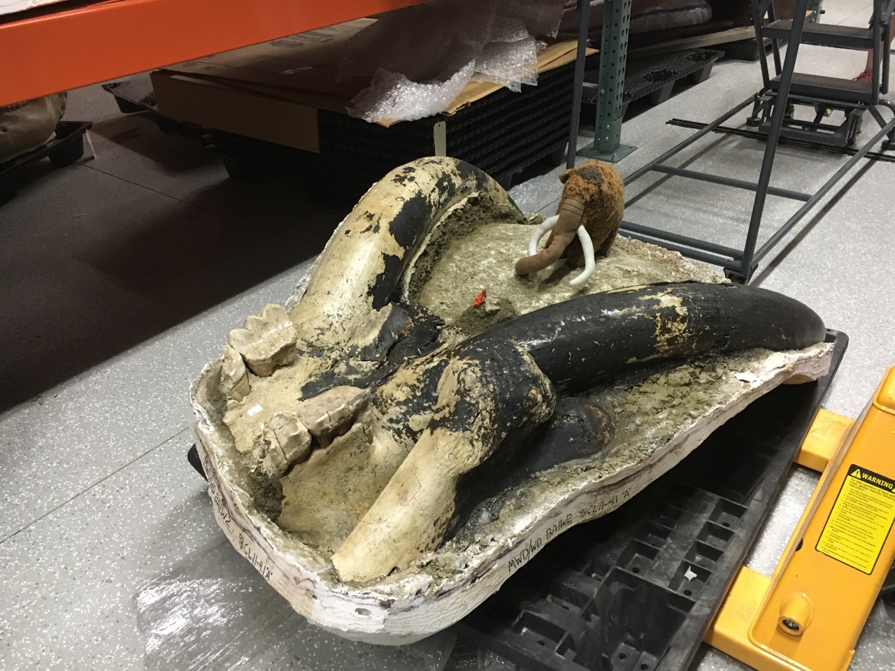 The proximal end of one femur is also preserved; it's the lump of bone in the foreground.The most notable thing about this skull is the black coloring over much of the surface, including almost all of the left tusk. It appears that this specimen was burned, almost certainly after death. We see this in a fair number of bones from Diamond Valley Lake, but it's the first time we've noted it in a mastodon, leading our Marketing Specialist Brittney Stoneburg to nickname this specimen "Blaze".Blaze is missing most of his third molars, but his second molars are intact and we were able to get measurements for the Mastodons of Unusual Size Project. Katy was also able to get measurements for her studies on mastodon tusks (and, based on the size of the tusks, Blaze was almost certainly a male). Blaze's second molars were heavily worn, and the anterior portions of his third molars were in wear, suggesting that he was close to the same age as Max when he died, perhaps in his mid-30s.Today on World Elephant Day, help support elephant conservation by visiting or donating to a museum, zoo, or conservation group that furthers our knowledge and protection of these amazing animals.
Fossil Friday - associated mastodon material
 This week we have two visiting researchers at Western Science Center. Dr. Katy Smith from Georgia Southern University has been measuring and photographing the proboscidean tusks in our collection, which we hope will lead to all kinds of new information about southern California mastodons and mammoths. Dr. Bernard Means from Virginia Commonwealth University's Virtual Curation Lab has been here on a trip sponsored by Smithsonian Affiliations to make 3D scans of some of the WSC specimens (Bernard has written about his visit here). These visits have meant that we've been pulling out lots of specimens, many of which I had never seen before.Last June I wrote about one of our mastodon jaws, which is an interesting specimen for the Mastodons of Unusual Size project because of its outlier status. This mastodon has the widest lower third molars we've measured in any California specimen, by a pretty large margin. As it turns out (and unknown to me until this week), we have part of the cranium from the same specimen.In the image above, the cranium fragment is sitting beside the lower jaw, and facing the opposite direction, so anterior is to the left. The fragment consists mostly of the left premaxilla and maxilla with the associated teeth. The rather large left tusk suggests that this was a male mastodon (the distal end of the tusk is broken off). The left second and third molars are both well preserved, with the second molar fully in wear and the third molar just starting to erupt, suggesting he was in his early 20's (and consistent with the lower jaw tooth wear).It turns out the upper third molars are outliers, but not quite to the same degree as the lower molars. We have several California upper M3s that are wider than this tooth, but those teeth are also much longer than this one. That means that the proportions of this tooth are unusual for California, even though we have teeth that are both longer and wider than this one. In fact, there's a tooth in our database from Ohio that is almost exactly the same dimensions as this one.
Fossil Friday - Isorthoceras sociale
 During the Mastodons of Unusual Size road trip, Brett and I made several stops to collect fossils for WSC. Since I was 20 years old, I've made several visits to the small, well-known (to paleontologists) roadcut in Graf, Iowa (the photo below was from a trip I made there in 2007):
 The rocks at Graf are from the Late Ordovician Maquoketa Formation, about 445 million years old. A number of different types of marine invertebrate fossils are found here, but perhaps the most prominent is the cephalopod Isorthoceras sociale.Cephalopoda is the group of mollusks that includes modern squid, octopus, and cuttlefish. Another group of modern cephalopods are the nautiloids, such as the chambered nautilus:
 Unlike other living cephalopods, nautiloids have an external shell made of the mineral aragonite. The shell in modern nautiloids is shaped like a coiled cone, and is divided internally into chambers:
 While nautiloids are relatively uncommon today, for hundreds of millions of years they were among the most abundant animals in the ocean, and it was during the Ordovician Period that they first became widespread. While the living nautilus has a coiled shell, nautiloid shell forms were more diverse in the past. Many of the early nautiloids had straight, conical shells. At Graf, there are several rock layers that are made up almost entirely of the shells of the small straight-shelled nautiloid Isorthoceras, at least four of which are visible in the photo at the top of the page. Here are some reconstructions of what Isorthoceras may have looked like:
 Most of the shells in any given layer at Graf are aligned in the same direction, so from certain angles the shells are mostly seen in cross section:
 The shells also sometime erode out of the rock, and can be found as isolated fossils:
 During our short stop at Graf, Brett and I filled a 5-gallon bucket with rocks containing Isorthoceras and other Ordovician marine fossils which will be added to the WSC collections.
Fossil Friday - Annularia
 I've spent the last week trying to catch up on administrative work while pouring over all the data we gathered during our "Mastodons of Unusual Size" road trip. But after several weeks of almost all mastodons it gives me the chance to feature a different organism for Fossil Friday. The specimen shown above was donated to the Western Science Center by the Earlham College Geology Department, and comes from the famous Carboniferous Period deposits at Mazon Creek in Illinois. While this looks somewhat like a flower, it's actually a whorl of leaves known as Annularia (flowers had not yet evolved in the Carboniferous). Annularia are the leaves of horsetails or scouring rushes, a plant that's still around as the genus Equisetum:
 Modern horsetails are small, spore-bearing plants that live primarily in marshy areas. But in the Carboniferous there were giant horsetails that grew to be up to 10 meters tall, and were among the world's most common trees.The name Annularia illustrates a particular difficulty faced by paleobotanists. If you're trying to identify a fossil vertebrate, you face several difficulties related to variability. Bones from different parts of the body look different, and they also vary with age (ontogenetic variation) and sex (sexual dimorphism). Plants present all these same issues, but they may also only grow some structures at certain times of the year (flowers, seeds, spores, pollen), the body parts may easily or even intentionally separate from the rest of the plant (deciduous leaves, seeds, spores, pollen), and preservation potential may be wildly different from one part to another. The result is that, if you have a bunch of fossil leaves and fossil stems, it's often very difficult to tell which ones go together unless that happen to be preserved actually connected to one another. Paleobotanists get around this problem by naming form taxa or organ taxa. The name Annularia specifically refers to leaf whorls of horsetails that share a particular morphology. Other parts of the plant, such as the stems and the spore cones, have different names. Fossil horsetails are quite common and well-known in Carboniferous rocks, so we actually know that fossil stems called Calamites come from the same plants that produce Annularia.
Mastodons of Unusual Size - Denver Museum of Nature and Science
 On Thursday Brett, Max, and I made our sixth stop on the "Mastodons of Unusual Size" tour, at the Denver Museum of Nature and Science.The major reason for stopping in Denver was to examine specimens from the Snowmastodon Project. In 2010 during the construction of a reservoir at Snowmass Village several thousand Ice Age fossils were discovered. There were two especially remarkable features at the site. First, at an altitude of over 8,800 feet, it was one of the highest Ice Age deposits ever found. Second, by far the most common animals were mastodons, accounting for more than half of the bones recovered. From the Rocky Mountains west to the Pacific Ocean, the vast majority of known mastodons come from just three sites - Snowmass Village, Rancho La Brea, and Diamond Valkey Lake.There are a number of very well-preserved crania and mandibles from Snowmass in the Denver collection (Max is seen above with some of the mandibles), that range from very young, perhaps even fetal, animals to an individual that was so old that its last tooth was worn to the roots (we couldn't measure that one because there wasn't enough tooth left!):
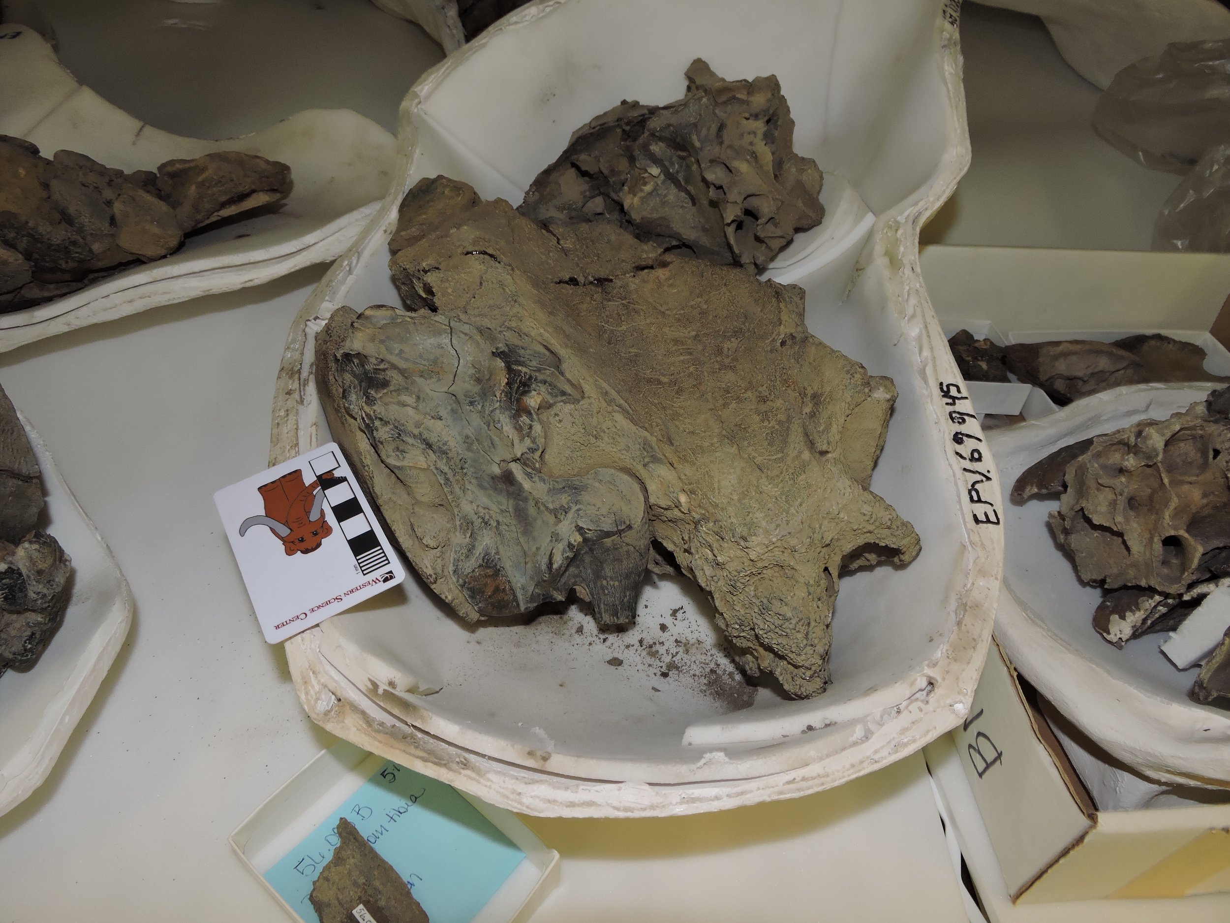 With such a large mastodon sample, we were able to measure teeth from every position and a variety of different ages. This tiny maxilla fragment preserves the first two teeth, the second and third premolars:
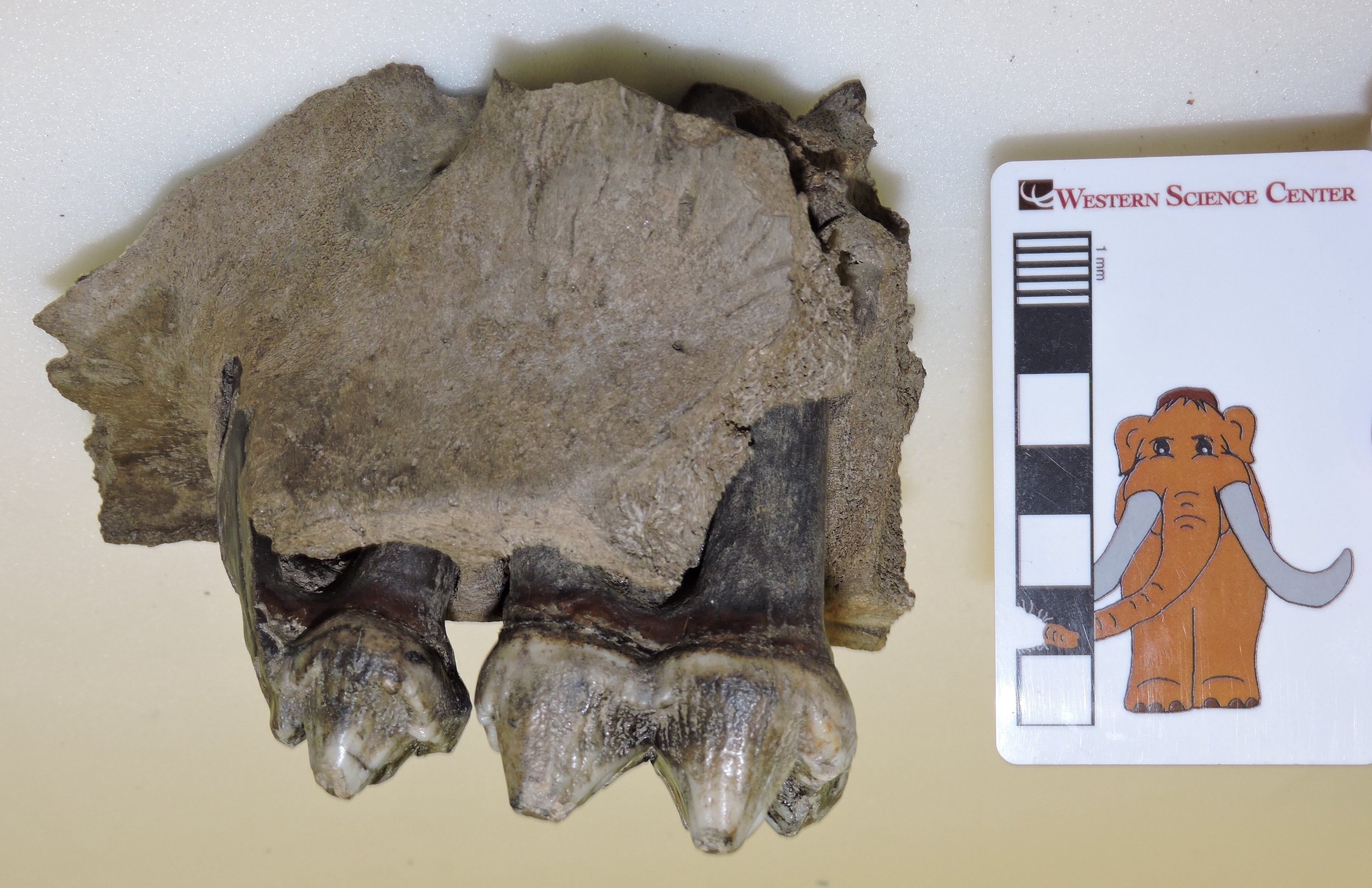 This maxilla has the third and fourth premolars:
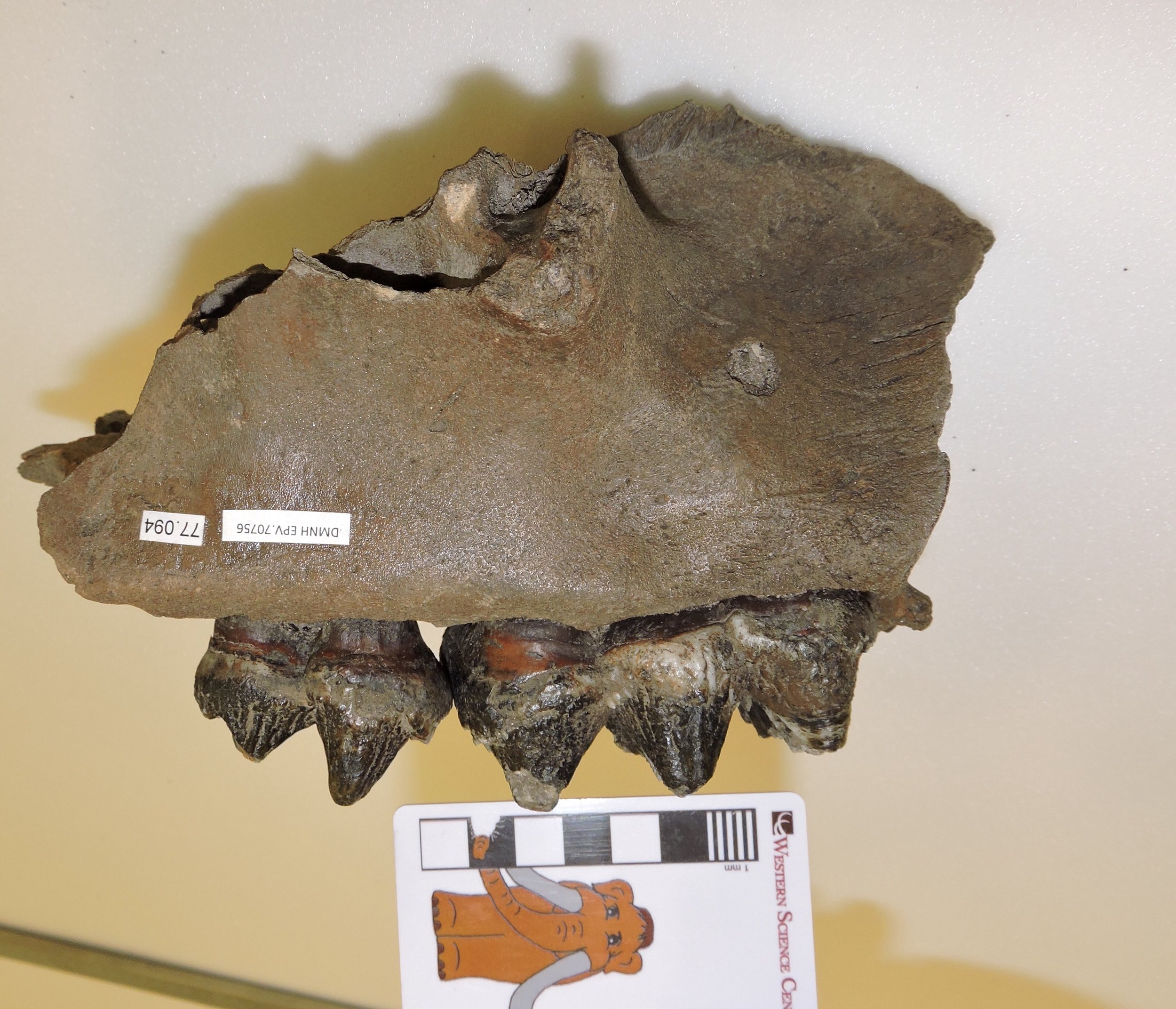 And below is a maxilla fragment with the next three teeth, the first, second, and third molars all preserved:
 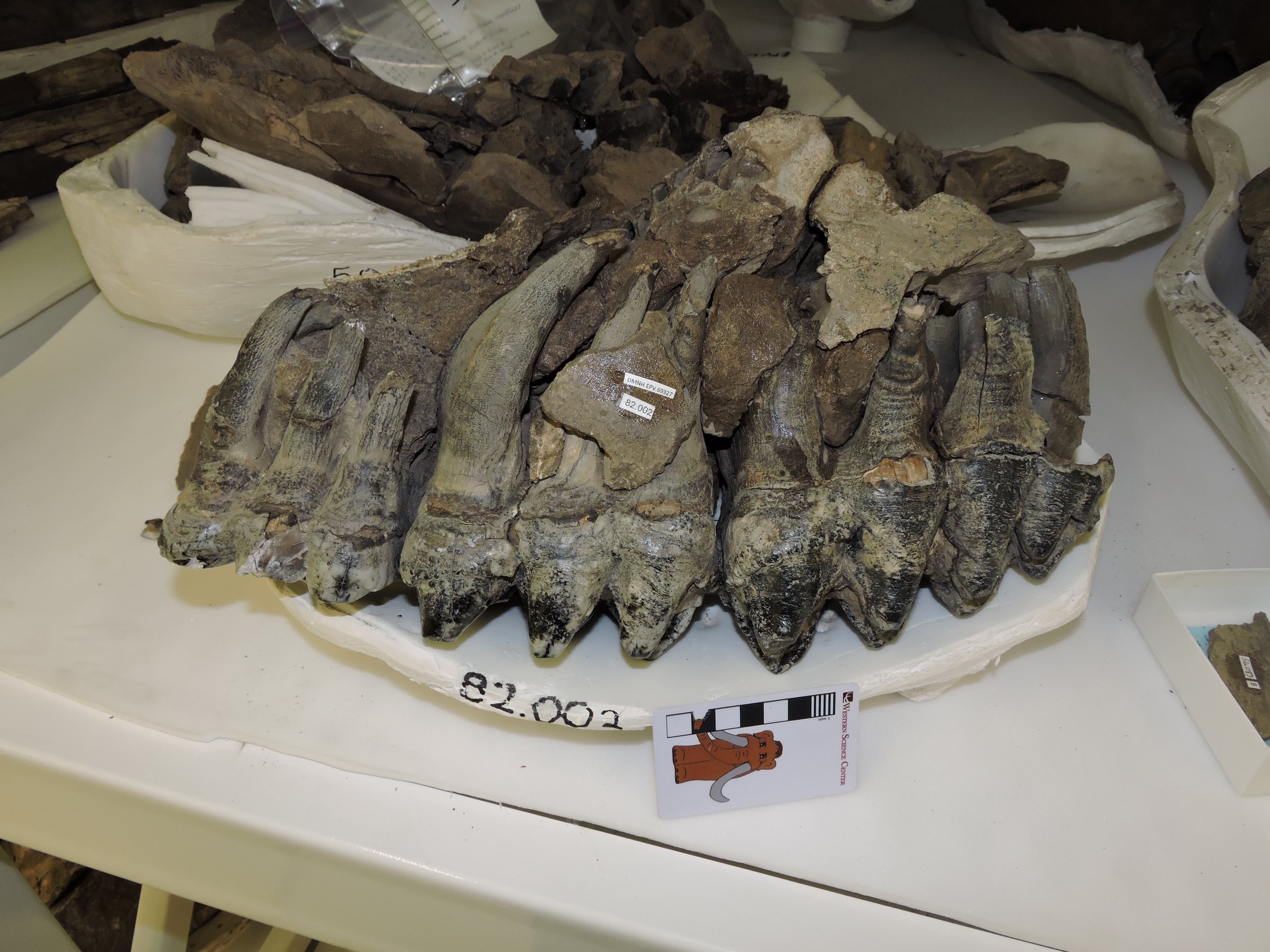 Some of the Snowmass specimens are true giants. We measured one femur (actually the cast, below) that had a distal width 11% greater than Max's:
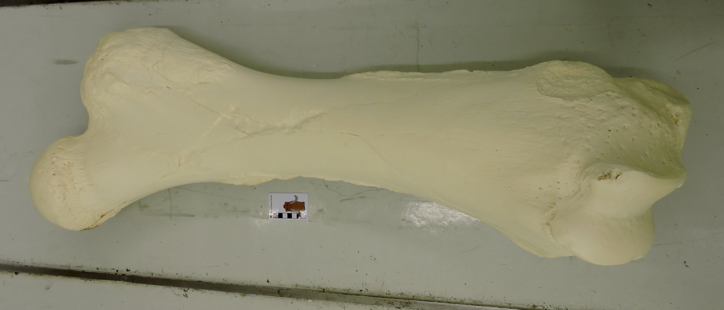 The Snowmass sample was an excellent addition to our dataset, but there were actually a few other mastodons in the DMNH collections. Below is apparently the only known mastodon tooth from Colorado that wasn't found at Snowmass:
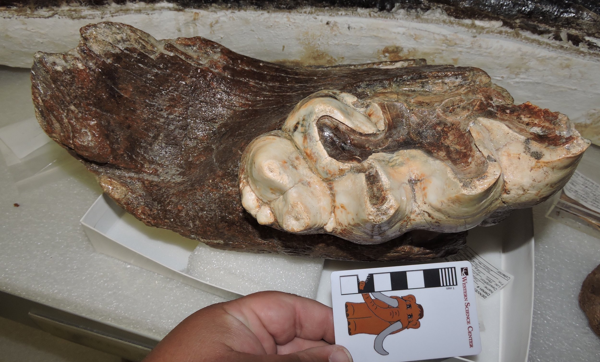 There is also an Indiana mastodon in the Denver collections. This specimen has femora that are slightly larger than Max's, and relatively rare 5-lophid lower third molars:
 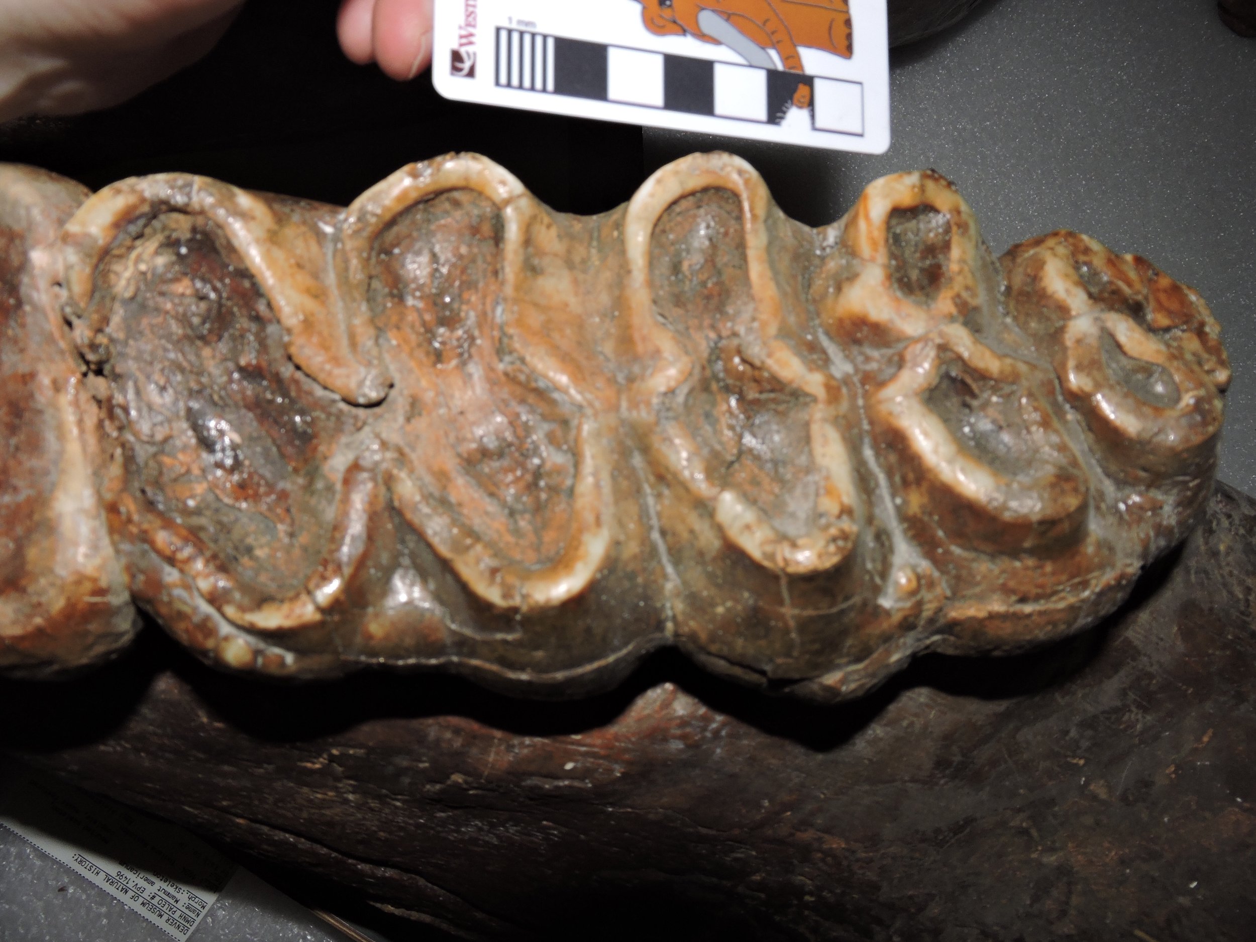 We spent seven straight hours in the Denver collections, measuring 85 mastodon teeth. It's going to take awhile for me to sort through everything and see what we have, but a few initial impressions did jump out at me. Most significantly, the Snowmass mastodons don't seem to look much like the California mastodons. Almost none of the third molars have a length:width ratio of greater than 2 (I think only one specimen), while almost every California mastodon has a ratio greater than 2. More strikingly, it seems that every Snowmass mastodon had lower tusks (as seen below), a feature that has never been observed in a California mastodon.
 As I write this, Brett, Max, and I are on our way home to California. But this doesn't mark the end of "Mastodons of Unusual Size". We are still gathering data that's being supplied by other researchers, and still collecting data on California mastodons (we haven't even measured all the Diamond Valley Lake specimens yet). In October we'll be presenting our preliminary results at the Society of Vertebrate Paleontology meeting in Salt Lake City. And, of course, I'll be posing updates on our progress here, on Twitter, and at experiment.com.Thanks to all the curators and collections managers who provided access to the collections in their care, and to our experiment.com donors that made this trip possible.
Fossil Friday - mastodon astragalus
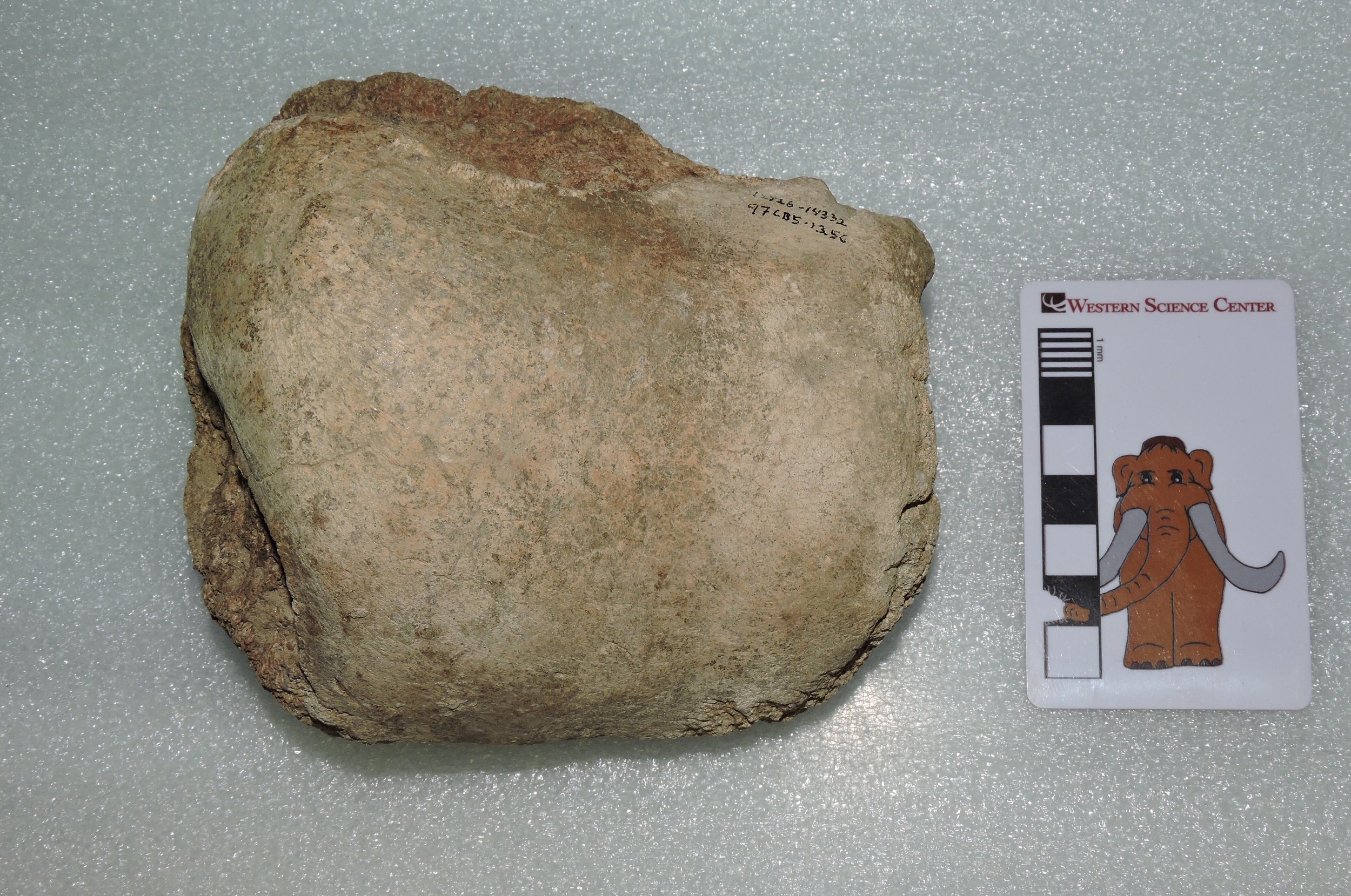 I'm still on the experiment.com-funded Mastodons of Unusual Size tour, and will be doing an additional post on that tomorrow. My inability to type and drive at the same time means I can only do a short Fossil Friday post.The bone shown above is a mastodon astragalus, the most proximal of the ankle bones. The upper end of the astragalus articulates with the lower ends of the shin bones (the tibia and fibula), although in elephants the amount of movement at that joint is limited. Proboscidean ankle bones are much less complex than in most other animals, and are optimized for weight-bearing. Interestingly, it seems that some dwarfed elephants evolved a rather different ankle structure for walking in hilly terrain.This astragalus was found at the East Dam of Diamond Valley Lake.