

 Tomorrow the United States celebrates Independence Day, commemorating the signing in 1776 of the Declaration of Independence. The primary author of the Declaration was Thomas Jefferson, who went on to hold various U.S. government office including vice-president and president. But in addition to his role in the founding of the United States, Jefferson played a major part in the development of paleontology in North America.In 1797, while he was vice-president, Jefferson presented a paper to the American Philosophical Society (published in 1799) reporting on bones that had been found in a cave in Virginia (now in West Virginia):
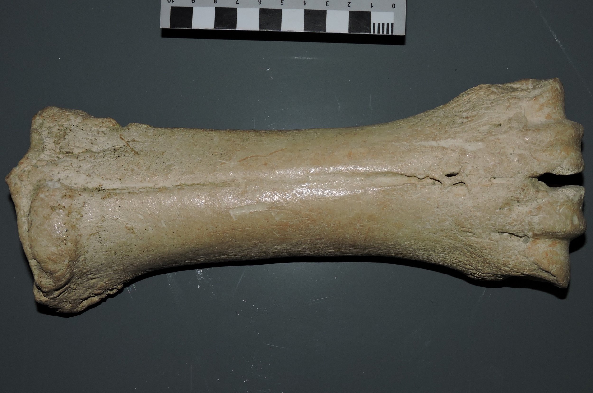
 Only a small number of bones were recovered, but these included three huge claws. Jefferson believed that the bones represented a type of giant lion, and proposed the name Megalonyx ("giant claw") for the newly discovered animal. It turned out that Jefferson's identification was incorrect. Two years later, Caspar Wistar correctly identified the bones as coming from a ground sloth. In 1822, A. G. Desmarest established the specific name jeffersonii for this species.While fossils from the Americas had of course been discovered and even figured in publications as early as the late 1600’s, very little scientific work on American fossils was published through the 1700’s. This changed starting in the early 1800’s, and Thomas Jefferson's publication of Megalonyx is often taken as marking the beginning of vertebrate paleontology in North America.Besides its historical importance, Megalonyx is a really interesting animal in its own right. It's a medium-sized ground sloth, up to about 3 m in length (some ground sloths were huge!). It's only distantly related to most other famous ground sloths such as Eremotherium and Paramylodon, and is in fact more closely related to the extant two-toed tree sloth Choloepus. Megalonyx was also the most wide-ranging of the ground sloths; while their remains are most common in the eastern U.S. they have been found from Florida to Alaska. A few specimens are also known from California, including a handful of bones from Diamond Valley Lake that are on public display at the Western Science Center. At the top of the page is a mandible, shown in dorsal view, with the same bone shown below in left lateral view:
  Only eight Megalonyx bones were recovered from the Diamond Valley and Domenigoni Valley excavations, but this still shows that Jefferson's ground sloth was roaming across California during the Pleistocene.References:Desmarest, A. G. 1822. Mammalogie, ou, description des espèces de mammifères, Chez Mme. Vueve Agasse, imprimeur-libaire, Paris.Jefferson, T. 1799. A memoir on the discovery of certain bones of a quadruped of the clawed kind in the western parts of Virginia. Transactions of the American Philosophical Society, 4:246-260.Wistar, C. 1799. A description of the bones deposited, by the President, in the museum of the society, and represented in the annexed plates. Transactions of the American Philosophical Society, p. 526-531.
Western Science Center theropod invasion?
 I arrived at work this morning to find what appeared to be several muddy tracks in the museum parking lot. While they weren't arranged in an organized trackway, they were numerous.
  These look a lot like small theropod dinosaur tracks, complete with claw impressions, such as this one from Dinosaur State Park in Connecticut:
 Of course, my first thought was that the museum was under attack from a pack of angry, ravenous theropod dinosaurs, presumably seeking revenge for my disparaging comments about the Jurassic World "Velociraptors". Upon further reflection and observation, it turned out that there was a more mundane explanation. The museum has several sweetgum trees around the parking lot. These leaves are generally five-pointed (below), but when the leaves fall and dry out in the sun the edges tend to curl up.
 We've actually had a little rain in Hemet the last few days, and the ground is a bit muddy. The leaves apparently got muddy on one side, then blew into the parking lot overnight. Then, before morning, the leaves blew away, leaving a three-point mud outline (the curled-up lateral portions of the leaves apparently didn't touch the ground).As a result, I was able to make it safely to my office, free of any Velociraptor-related unpleasantness.
Fossil Friday - wildfires
 A large wildfire, called the "Lake Fire", is currently burning in the San Bernardino National Forest. Even though the fire is about 50 km north of Hemet, smoke from the fire is clearly visible from the Western Science Center.Wildfires such as this are widespread in Southern California during the summer, driven by dry vegetation and frequent afternoon and evening winds. The regularity of these fires is evident in the fleet of Cal Fire aerial tankers based at the Hemet Airport, which are currently making regular flights to combat the Lake Fire:
 Anthropogenic climate change and the ongoing drought have resulted in ideal conditions for wildfires in this area, but wildfires are not a new occurrence in California. In fact, a quick records search of the Diamond Valley Lake fossil collection housed at the museum turned up at least 80 Pleistocene specimens that show evidence of burning. The DVL fossils all predate the arrival of humans in California, so these aren't animals that were cooked for food. They represent animals that were exposed to wildfire at or fairly close to the time of death.In some cases the evidence for burning is subtle, but in others there is no room for doubt:
 Above are two views of the left tibia (shin bone) of the camel Camelops. Most of the bone is missing, with only the distal end preserved (there is also a box full of associated fragments). The bone is completely burned, inside and out, and has almost a charcoal-like texture on the surface. The burning extends to the broken surface, so presumably the bone was broken when it burned. It's possible that the bone had been exposed on the ground for awhile and had already started cracking up when the fire came through, but it's also possible that a relatively fresh bone cracked and broke due to the heat from the fire. Regardless, specimens like this show that, much like today, wildfires were a regular occurrence in Southern California during the Pleistocene.
Fossil Friday - bison metacarpal
 This week's Fossil Friday features a metacarpal (hand bone) from a Pleistocene bison. The example shown here is a left metacarpal shown in anterior view, with the proximal end (at the wrist) on the left and the distal end (at the first knuckles) on the right.Like many other advanced artiodactyls, in bison this structure is actually two bones that are fused together. These are the third and fourth metacarpals, the hand bones that in humans support the middle finger (Digit III) and the ring finger (Digit IV). Both of these "fingers" are present in bison as well, with each supporting a hoof (remember that our hands are just highly modified front feet). The articulations for each digit are visible at the distal end of the bone, on the right side of the picture.Here's the same bone in posterior view (the equivalent of "palmar view" in a human hand):
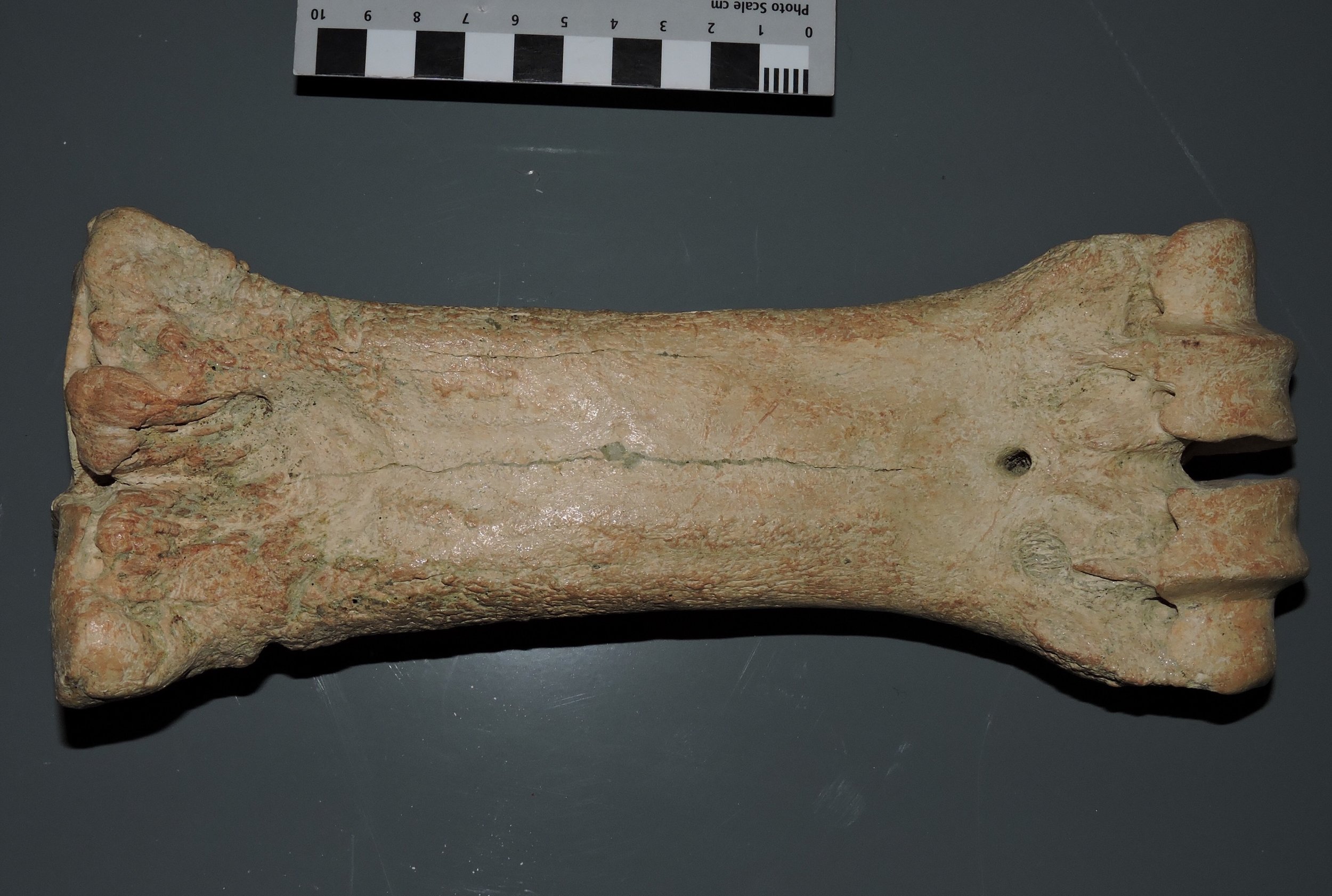 In this view the articulations for digits III and IV are more clearly visible on the right, but the groove indicating the line of fusion between the third and fourth metacarpals is almost completely lost.Last year I posted photos of the metacarpal from the extinct camel Camelops. Camels and bison are not very closely related to each other, but they are both artiodactyls that show similarities is the hand and foot structure. In both animals the front foot is reduced to just the fused third and fourth metacarpals. Camelops was a large animal that was much taller than Bison, and this is reflected by the relatively long, slender metacarpal. Bison, while shorter, were heavier and had to support a massive head, resulting in a shorter, stouter metacarpal.
Fossil Friday - eagle talon (or, a Pleistocene dinosaur)
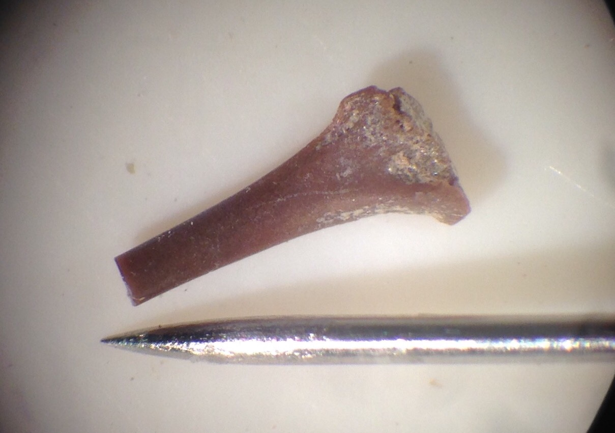
 With the release of Jurassic World, we've been getting some inquiries from the media about what dinosaurs the Western Science Center has in our collections. While we have a small number of isolated bones and teeth from the Cretaceous Hell Creek Formation in Montana, the answer that they really don't like to hear is that we have lots of dinosaurs, because we have lots of birds.In general, the paleontological community's reaction to Jurassic World has been disappointment at best (disclosure: I haven't seen the movie, and have no immediate plans to do so). The problem is that, while the original Jurassic Park movie had a lot of errors, it did a pretty good job of portraying dinosaurs based on our scientific knowledge at the time. But we've learned a lot more in the 20+ years since, and Jurassic World doesn't reflect that; in fact, in some ways it seems to be a huge step backwards even relative to Jurassic Park. One of the key points where Jurassic World fails is in it's failure to use birds as a model for reconstructing dinosaurs. Whether or not the Jurassic World designers like to admit it, birds are the last surviving branch of dinosaurs. They also happen to be a highly successful branch; there are roughly twice as many modern species of birds as there are of mammals. Even though birds have a relatively low preservation potential due to their fragile bones, with such a successful and widespread group we would expect them to show up fairly commonly as fossils. In fact, we have quite a few species of Pleistocene birds represented in the Diamond Valley Lake fauna. These are almost all based on isolated bones, such as the eagle talon shown at the top of the page. I'm not sure what species this talon represents, but a good possibility is a golden eagle, Aqulia chrysaetos (modern example below from the Nashville Zoo):
  Golden eagles were widespread in California during the Pleistocene; in fact, they are the most common animal at the Pleistocene tar pits at Rancho la Brea, and there are several specimens in the Diamond Valley Lake fauna. So, Jurassic World aside, California dinosaurs are alive and well!
Fossil Friday - partial horse skull
 Horses are among the most common large animals in the Diamond Valley Lake fauna, and there are several skulls in various states of preservation in the Western Science Center collection. One of these skulls stands out, however, because of the strange way in which it was preserved.The skull, which was collected at the East Dam of Diamond Valley Lake, is shown above in ventral view; essentially, you're looking at the roof of the mouth. While it has been mostly cleaned, it's still in the original field jacket, so this how the specimen was preserved in the ground. This view shows the complete set of posterior teeth (premolars and molars) from both sides of the upper jaw; the left ones are labeled below:
  You may have noticed that, while there are a lot of teeth in this jacket, there is no bone. Now, teeth tend to preserve better than bones, since tooth enamel is tougher than bone. But in this case we have the complete, well-preserved upper posterior dentition - 12 tooth positions - all in their correct positions relative to each other, with no bone to hold them in place! This suggests that the bone was removed after the skull was buried; otherwise the teeth would have shifted out of position. The only explanation I've been able to come up with is that, after burial, the skull was exposed to sediment or ground water chemistry that caused the bone to dissolve while leaving the teeth untouched. Differential dissolution rates are not unusual in fossils, but I don't recall previously seeing an example in which the bone was completely dissolved while the teeth were apparently untouched.This strange preservational history does provide us with certain advantages. With the bones removed, we can see features of the dentition that would normally require X-rays or CT-scans. Notice that M3 has a different appearance than the other teeth. That's because it had not yet erupted when this horse died, and was likely still imbedded in the upper jaw. This also enables us to estimate the horse's age. In modern horses M3 typically erupts when the horse is 3 to 4 years old, and this M3 was not even close to erupting, so this horse was almost certainly between 2 and 3 years old, likely closer to 2. That also means that its deciduous (milk) premolars should still be in the mouth, with the permanent premolars still developing. So let's look at a side view of the left premolars:
  Here's an annotated version:
 As expected, the permanent premolars were still developing when this horse died, and are present beneath the deciduous premolars (actually above them, since this is the upper jaw sitting upside down). Their presence also helps refine our age estimate, since P2 should erupt around age 2 to 3 years. Since dP2 is still in place but almost worn away, this horse was around 2 years old when it died.While in general I prefer to use X-rays or CT to look inside a bone, this skull gives us a great view of what was happening in this horse's dental development, and provides some interesting hints about the geochemistry of the sediments from this site.
Fossil Friday - Carboniferous mollusks
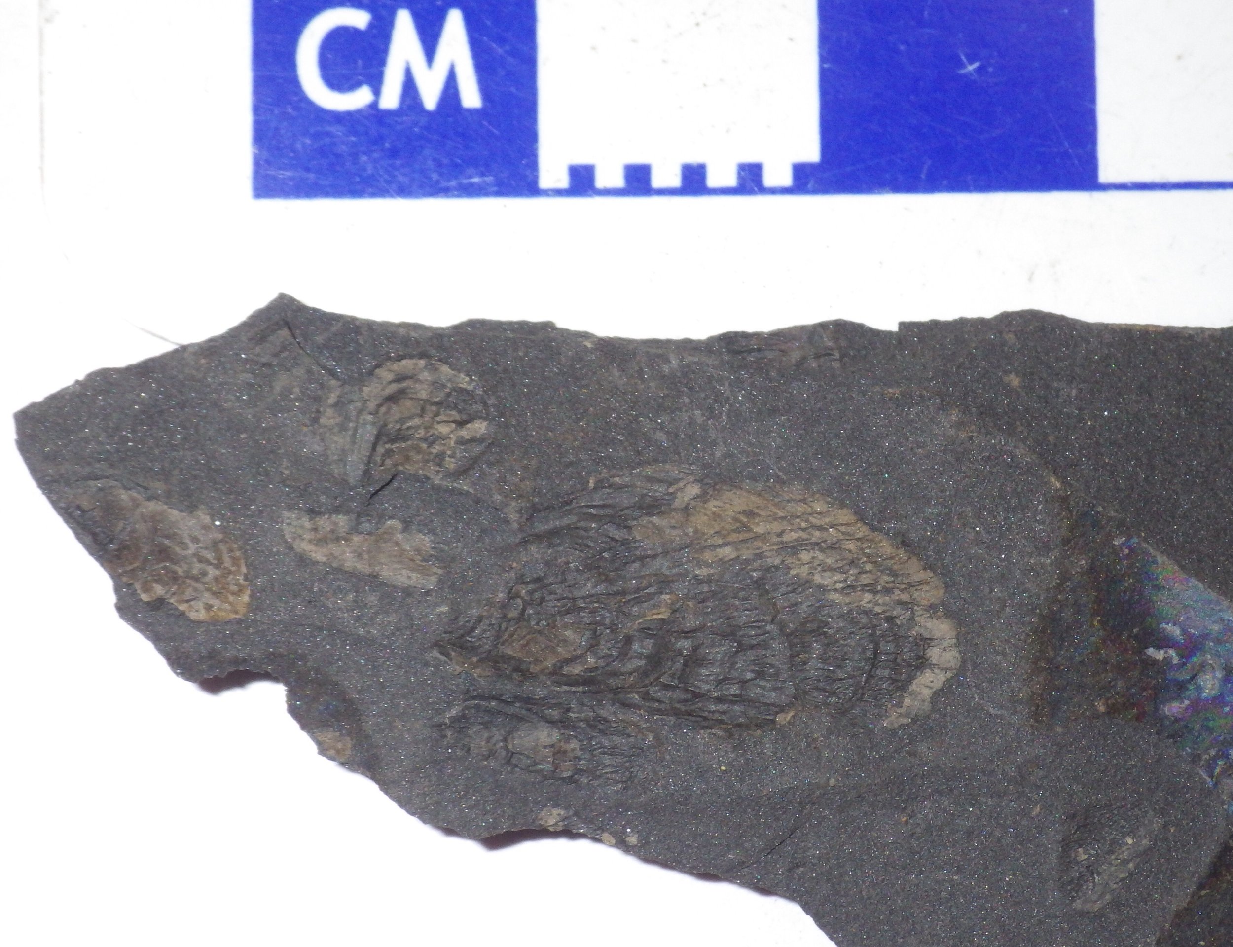 As Brett, Tim, and I headed back to Virginia after our fossil plant collecting trip to Beckley, West Virginia a couple of weeks ago, we made a second brief collecting stop in Princeton, WV. Several years ago, Tim and I stumbled onto a fossiliferous outcrop while fixing a flat tire of my field truck, so we revisited this site to collect some specimens for WSC.The outcrop exposes the Late Carboniferous Pride Shale Member of the Bluestone Formation, which seems to represent a shallow intertidal environment. It looks like all the fossils we collected are tiny pteroid mollusks, most of which are less than 2 mm long. We collected a few small pieces of shale (the outcrop is weathered, and the shale tends to fall apart), which contains perhaps 50-100 mollusk specimens. There is also a possibility that fossils of other organisms could turn up once we've had a chance to examine the rocks more carefully.
Fossil Friday - Carboniferous plants
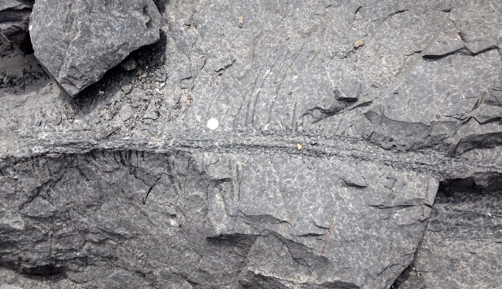 Last week I made a short trip back to Virginia for my son's graduation from Patrick Henry Community College. This also was a perfect opportunity for some fossil collecting, so Brett, Tim, and I met DorothyBelle Poli and Lisa Stoneman from Roanoke College for a day trip to Beckley, West Virginia.Boxley Materials operates a quarry near Beckley that cuts through the Carboniferous (~ 318 million-year-old) New River Formation. Certain beds of the New River Formation are full of fossil plants, and Boxley set aside some of this material for us to examine. Like many Carboniferous forests, one of the most prominent types of plants in these deposits is giant lycopods. Above is an example of Stigmaria, part of the root system of these lycopods (because it's rare to find all the parts of a plant in a single fossil, different fossil plant structures often have different scientific names). Unfortunately, this Stigmaria was in a rock that was far too large for us to collect.Giant lycopods are sometimes called "scale trees" because of the geometric (often diamond-shaped) leaf scars on their trunks:
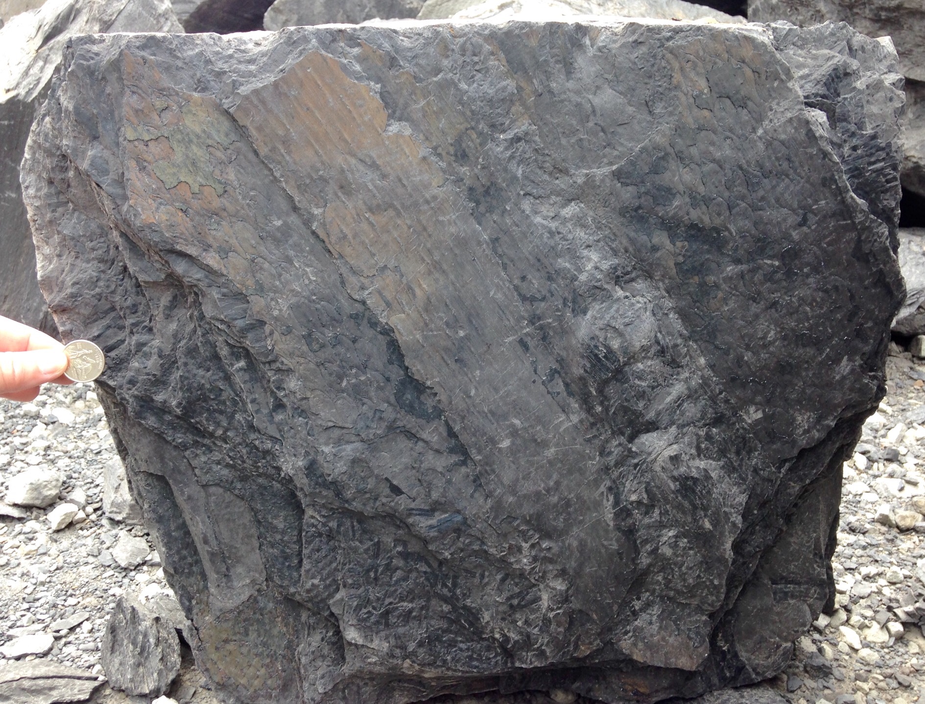 Seed ferns are also widespread in Carboniferous deposits; there are several different species in this sample:
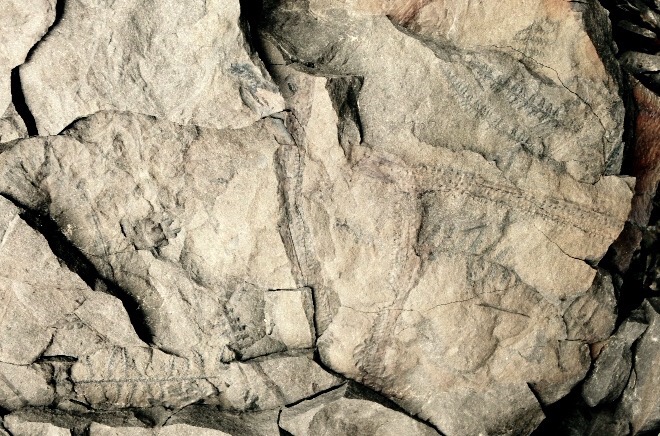 We are able to collect several good specimens for the Western Science Center, which will be nice additions to our growing paleobotany collection.
Fossil Friday - Bison atlas vertebra
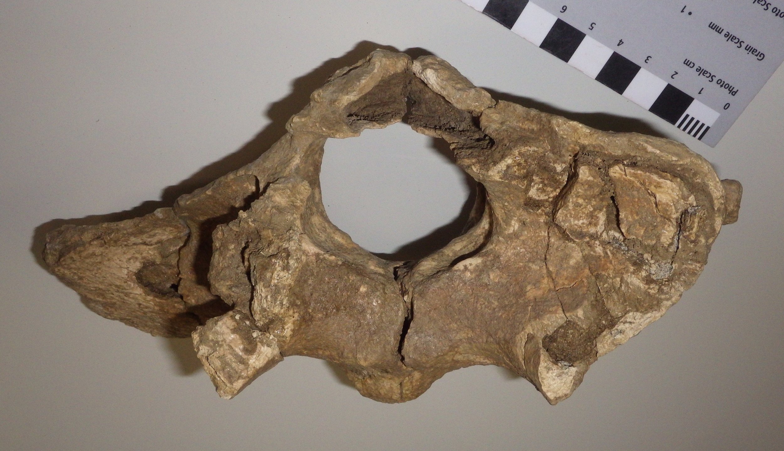 For this week's Fossil Friday I'm going to stick with bison, this time featuring an atlas vertebra.As I've described in earlier posts, the mammalian vertebral column is generally divided into five different regions. The most anterior are the cervical (neck) vertebrae. Almost all mammals have exactly seven cervical vertebrae, but mammals are unique among vertebrate classes in having such a tightly constrained cervical count. The first two cervical vertebrae, the atlas and the axis, are always highly specialized and look very different from any of the other vertebrae.That is mainly because of the complex articulations necessary to allow a range of movement in the head. When you nod your head (flexion and extension), that motion primarily occurs at the joint between the front of the atlas vertebra and the skull. When you shake your head (rotation) the motion is mostly at the joint between the back of the atlas vertebra and the front of the axis vertebra. That all means that the atlas has to accommodate one type of motion (nodding) on its front surface, a different motion (rotating) on its back surface, while providing attachment points for all the muscles that actually cause the movement, and allowing a passageway for the spinal cord and the vertebral arteries and veins through the middle of all of this movement. It's no wonder that the atlas has such a distinctive appearance. (As an interesting aside, whales, which have almost no ability to move their heads, still have complex and unique atlas and axis vertebrae, a relict of their ancestors that had much more mobile necks.)So, with all that said, the image at top shows a bison atlas vertebra in anterior (cranial) view. The bone is somewhat crushed and incomplete; much of the right side in particular is missing (remember, since this is anterior view, the right side of the vertebra is on the left side of the photo). The large hole in the bone is the neural canal, the passageway for the spinal cord. Below and beside the neural canal (centered at the "5-o'clock" and "7-o'clock" positions) are large depressions that articulate with the occipital condyles on the skull; this is the main "nodding" articulation.
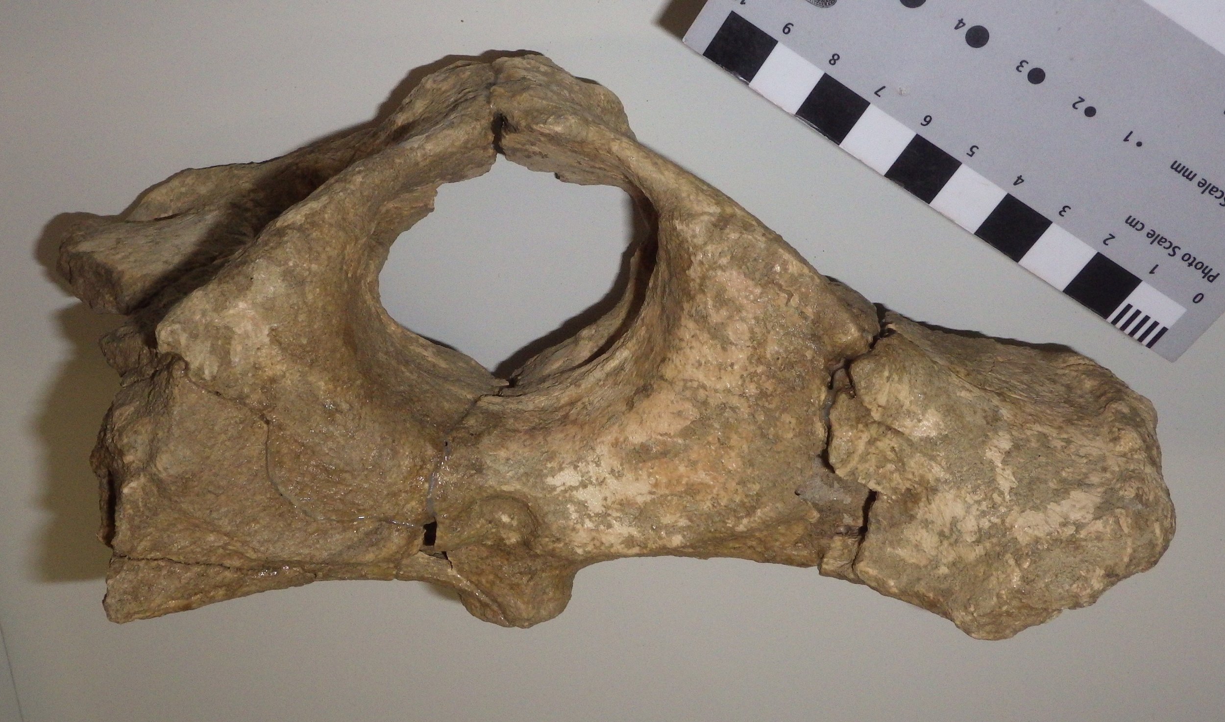 Looking at the same vertebra in posterior (caudal) view, we see broad, flat surfaces below and beside the neural canal. These are the articular surfaces for the axis vertebra, where rotation occurs. The large projection of bone on the right is a lateral or transverse process (the corresponding one on the left side is broken off). This provides a surface for the attachment of neck muscles associated with head movement. Bison have large, heavy heads, and so the muscle attachment areas to support them are relatively large.Here's the atlas in dorsal (top) view:
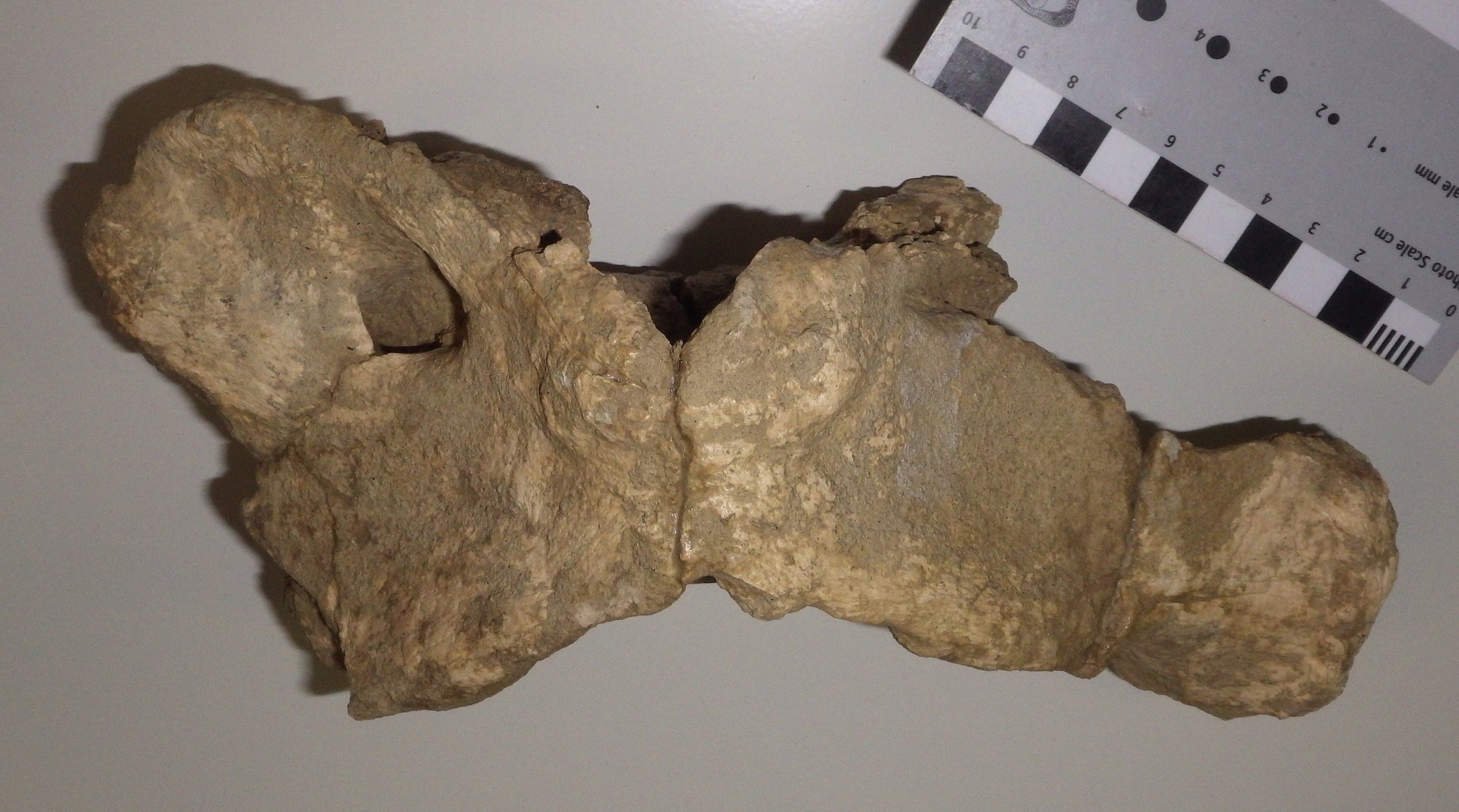 At this angle you can see that the transverse process is broad across the top surface, and has quite a large surface area. The opening toward the upper left is the alar foramen (the right one is only partially preserved), which provides a passageway for the vertebral arteries and veins.Even allowing for damage to the bone, the shape of this atlas seems a bit different from that of the modern Bison bison (see this site for an example). There are two different species of Bison known from the Diamond Valley Lake fauna, B. antiquus and B. latifrons. B. latifrons has a larger, heavier head than B. antiquus, and it's possible that this might be reflected in the anatomy of the cervical vertebrae, but I've not yet checked references to confirm this. At the moment, we have this particular atlas identified as Bison sp.
Fossil Friday - "Then the rats got him"
 Bison are among the most common large animals in the Pleistocene Diamond Valley Lake fauna, but like almost all the remains from these deposits they are usually fragmentary. But even fragmentary fossils can provide a lot of information, including the bison right lower jaw fragment shown here.Sometimes bonebeds or other rich deposits of fossils are formed during catastrophic events, with large numbers of organisms buried together very rapidly. That's not what we see in Diamond and Domenigoni Valleys, however; these deposits are primarily the result of the gradual accumulation of skeletons over thousands of years. That means that an individual skeleton may have laid exposed at the surface for a significant period of time before it was eventually buried. This exposes the bones to sun, wind, and rain, all of which can cause the bone to deteriorate and the skeleton to fall apart, and to scavengers, which may damage or eat the bones, or carry pieces away. All these things contribute to the fragmentary nature of the Diamond Valley Lake specimens.Evidence of the specific types postmortem trauma experienced by these bones is sometimes preserved. If we zoom in on the bison jaw shown above, we can see a series of grooves cut into the bone:
 These grooves are bite marks, most likely made by rodents. Bone gnawing has been documented in lots of different rodents, including rats, mice, and squirrels. It's generally attributed to the need for rodents to keep their ever-growing incisors from becoming too long, and perhaps to provide an additional source of calcium. But the presence of these marks immediately tells us quite a bit of additional information; the bison died in a setting that didn't result in immediate burial, and rodents were present in the environment.Of course, from the bison's point of view, maybe this is only adding insult to injury!
3D models of WSC specimens
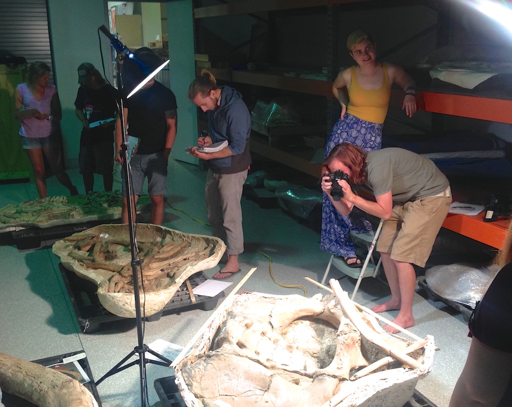 Last Saturday Pomona College's Karl Lang brought his geology class on a field trip to the Western Science Center, to learn about California's Pleistocene fauna and museum operations. While they were here, the students also took photos of several mastodon jackets in our collection in order to produce digital 3D models, which they've been kind enough to post online.Each image below has an embedded link to take you to a 3D viewer. Within the viewer, the images can be rotated and zoomed (I unfortunately couldn't get the viewer to embed properly in Wordpress).A partial vertebral column and ribcage:
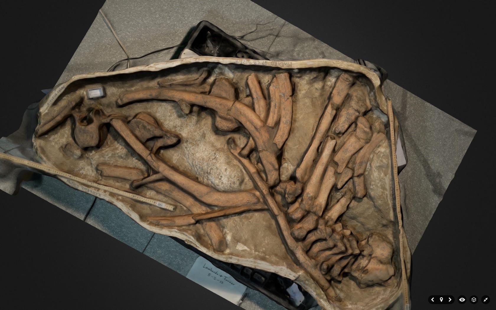 A pelvic girdle:
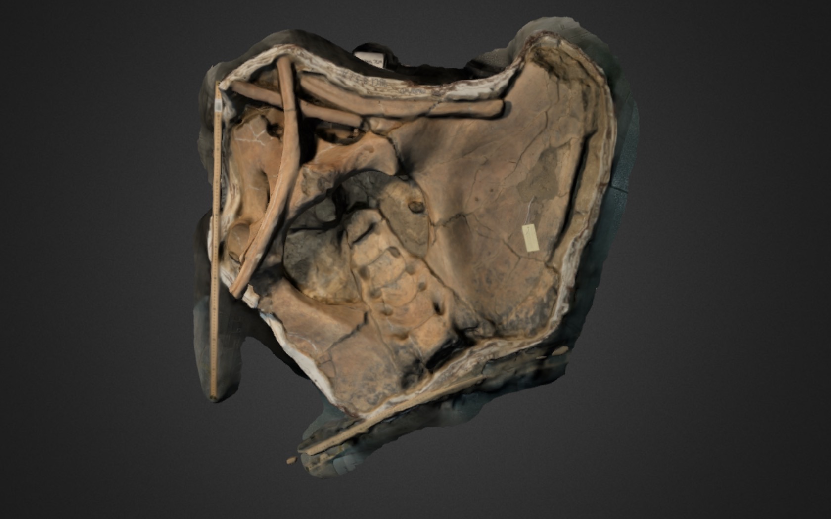 A partial skull:
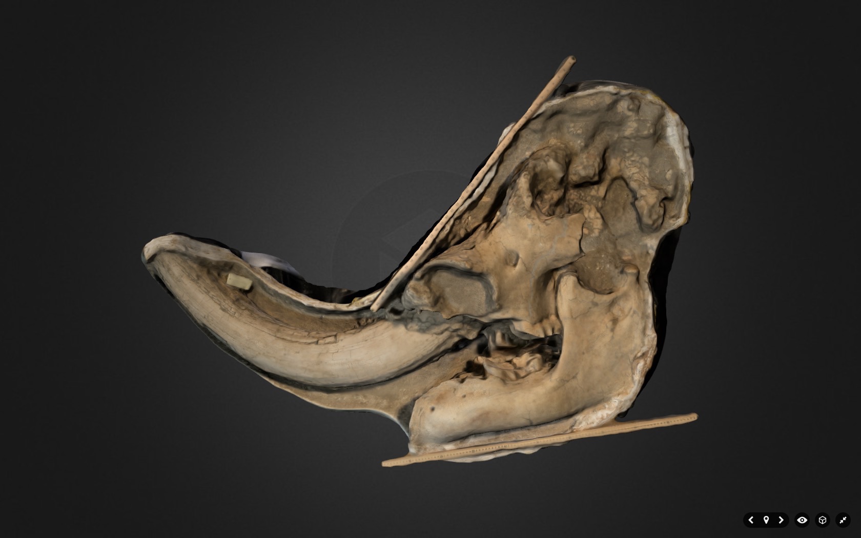 Thanks to Karl and Pamona's geology students for visiting, and for producing these great models!
Fossil Friday - western pond turtle costal plate
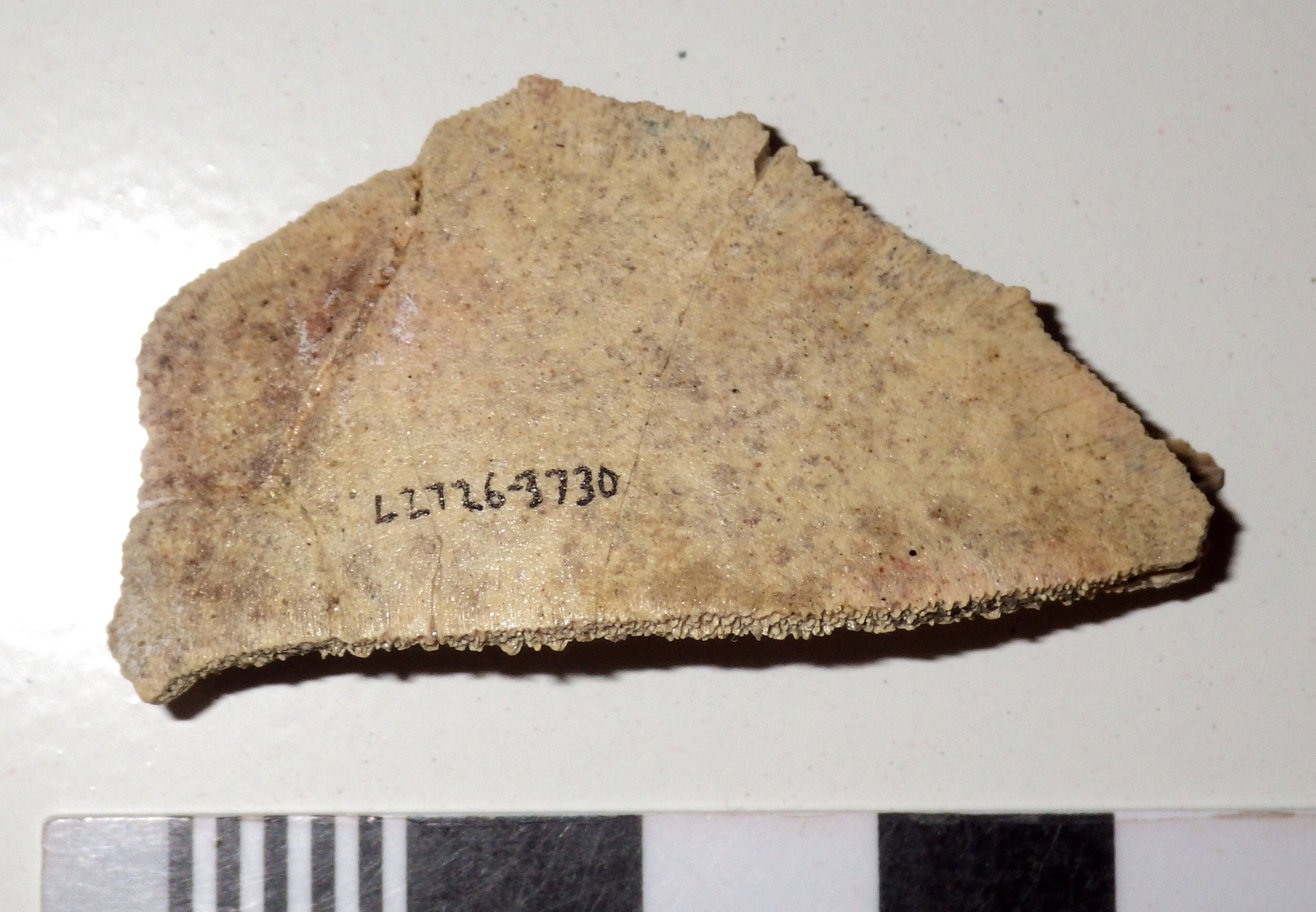 Fossil preservation is a tricky thing, and the variables involved in fossilization can have a major effect on what eventually gets preserved (a major subfield of paleontology, taphonomy, looks at this very issue). For vertebrates, one factor that can give you a high preservation potential is having a lot of bones, so that a large percentage of your body mass is more likely to survive. With their extensive bony shells, turtles should be a prime candidate for fossilization, and sure enough, large numbers were found in the Pleistocene Diamond Valley Lake fauna. Of course, having lots of bones is no guarantee that they'll survive in one piece; most of our turtle remains are fragments of the shell, and we have nothing even remotely approaching a complete turtle skeleton.Turtle shells have two main pieces, the carapace on the turtle's dorsal side and the plastron on the ventral side. The carapace and plastron, in turn, are each made up of numerous smaller bones. The piece shown above is from the carapace, technically called the first right costal, which formed the front right part of the shell. It's shown here in dorsal view; if the skeleton were complete, the turtle's skull would have been out of the image at the upper left. Here's a ventral view of the same bone (since this is from the carapace, it's like you're inside the turtle looking out):
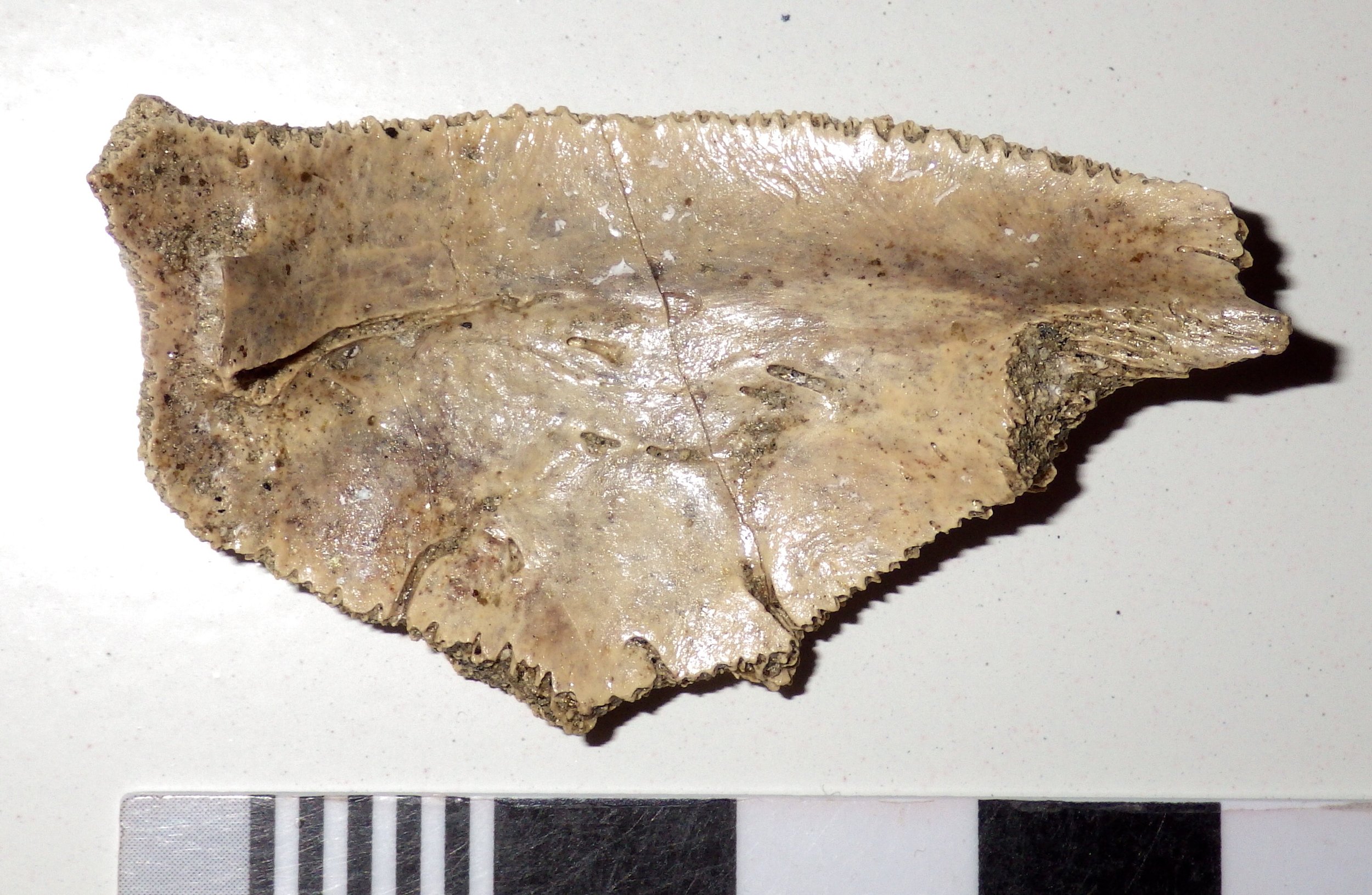 Most vertebrates that have bony armor (armadillos or crocodiles, for example) make their armor from dermal bones. These are bones that are embedded in the skin and generally have no direct connection to the rest of the skeleton. Many of the bones in a turtle's carapace are different in that they are attached directly to internal bones. The costal plates are attached to the ribs; this is more obvious in an oblique view of the ventral side of the bone, where the rib is clearly visible:
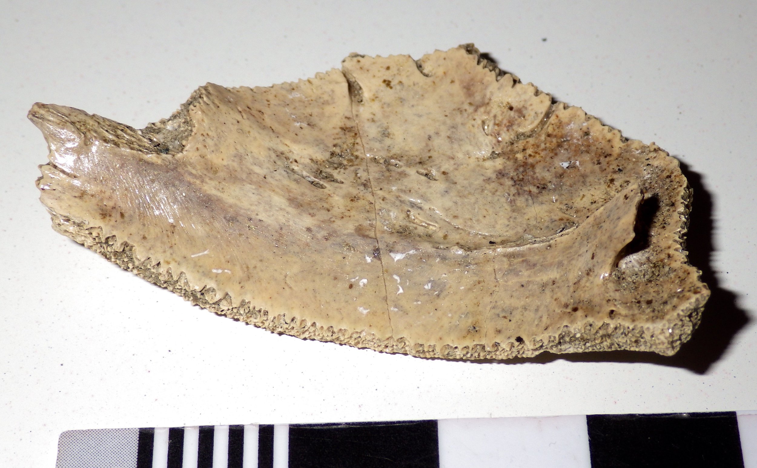 This particular costal bone is from a western pond turtle, Actinemys marmorota. These turtles are still found on the west coast, yet somehow I've never photographed one, even though I have dozens of turtle photos including every other genus in the family Emydidae. The closest relatives I've photographed are Blanding's turtle (Emydoidea blandingii) from the upper Midwest:
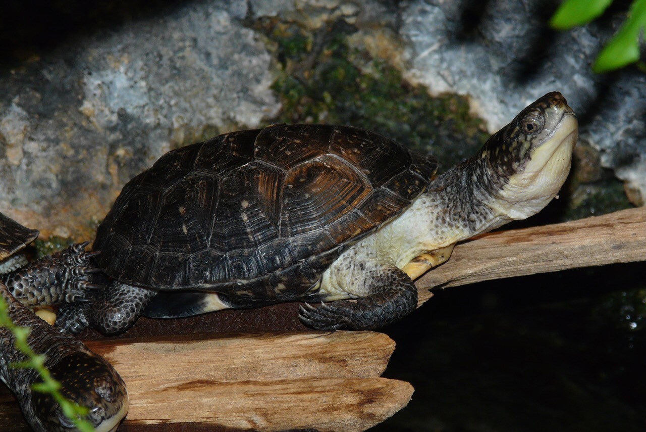 ...and the spotted turtle (Clemmys guttata) from the east coast:
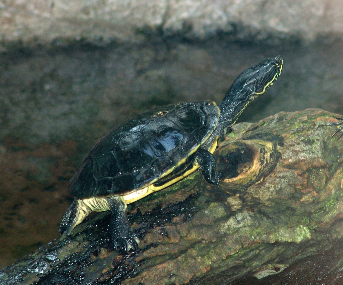 These three species are all closely related, and are in the same subfamily. In fact, until a few years ago Actinemys marmorota was placed in the genus Clemmys (our labels at WSC actually list these specimens as Clemmys marmorota); some workers have also proposed that Actimemys and Emydoidea in the same genus, Emys.
World Tapir Day
 World Tapir Day occurs on April 27 each year, to recognize and promote the conservation of tapirs. If you're unfamiliar with tapirs, they are perissodactyls (the mammal order that includes rhinos and horses) that currently live in South America (3 or 4 species) and Southeast Asia (1 species). Below is the Malayan tapir, Tapirus indicus, at the Henry Doorly Zoo:
 And here's a South American species, Baird's tapir (Tapirus bairdii) from the Jacksonville Zoo:
 Like their relatives the horses and the rhinos, much of tapirs' evolutionary history occurred in North America, and they only became extinct on this continent near the end of the Pleistocene. Water-loving tapirs apparently were not especially common in Southern California, at least not during the Pleistocene. None have been identified from the Diamond Valley Lake collections, and only a handful of tapir bones have been reported from Rancho la Brea. In the Western Science Center collections we only have a single specimen of a tapir (as far as we know), the four teeth shown at the top of the page (and a few associated tiny bone fragments) from near Temecula.All four teeth appear to be from the lower jaws, two from the left and two from the right. In the image I've oriented them they way I think they were located in the jaws, with the front of the jaw to the right. Without a modern tapir jaw on hand for comparison my identification is tentative, and I could be off by one tooth position, but I think the two left teeth (at the top) are the second and third molars (m2 and m3) while the right teeth (at the bottom) are the first and second molars (m1 and m2).It's not a lot, but these teeth demonstrate that tapirs were present in Riverside County during the Pleistocene. Happy World Tapir Day!
Fossil Friday - Harlan's ground sloth femur
 There are three different species of ground sloths known from the Pleistocene Diamond Valley Lake fauna. By far the most common of the three is Harlan's ground sloth, Paramylodon harlani. For this week's Fossil Friday we have a Paramylodon femur.This is the right femur, seen from the back (posterior view), with the distal end on the right. The articular surfaces for the knee joint are visible on the right, but the proximal end of the bone is not preserved so the ball joint where the femur attaches to the pelvis is missing (it would have been at the lower left corner of this image). Even though the bone is crushed, it has maintained its shape pretty well. Like many of the larger bones in the WSC collection, this one has only been prepared on one side.I've always found sloth femora fascinating, because they have a really strange shape when compared to most of the other mammals we find in Pleistocene deposits in North America. They appear to be very wide for their length, and generally kind of overbuilt for their size. To get an idea, compare this femur to the juvenile mastodon femur I wrote about a few months ago. These animals were probably roughly the same size, but the difference in their femoral proportions is striking.———On another note, we are busily preparing for tomorrow's Inland Empire Science Festival. If you're in Southern California we hope you can make it out to the Western Science Center tomorrow for this exciting event.
Fossil Friday - iguanid humerus
 In terms of numbers, most of the Pleistocene fossils from Diamond Valley Lake are from small animals, especially rodents and other small mammals. But there are non-mammals represented as well, such as the rattlesnake I posted a few weeks ago, and today's example, a lizard.This specimen is the proximal part of the left humerus, the upper arm bone. It has been identified as coming from the Family Iguanidae. The most famous iguanid is Iguana itself, but the family is fairly diverse with a number of different genera. Two species still live in Southern California, the desert iguana Dipsosaurus dorsalis:
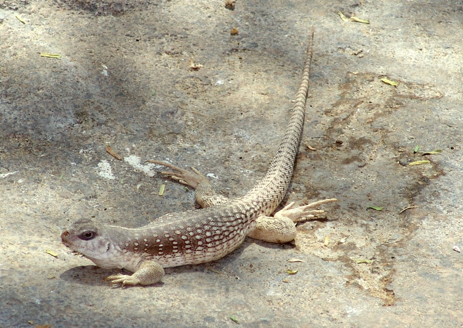 ... and the common chuckwalla Sauromalus ater:
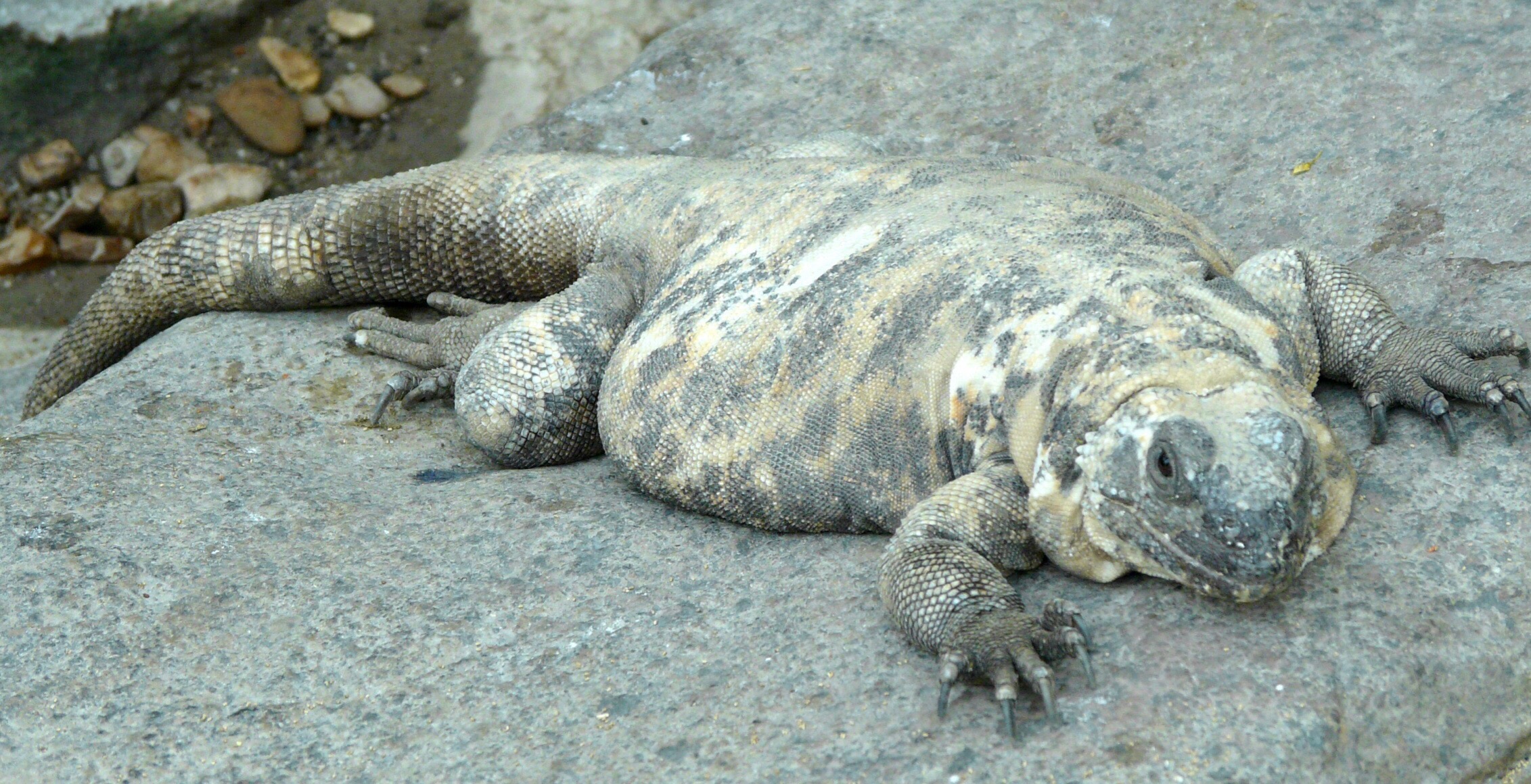 There is also an extinct iguana from Riverside County, Pumila novaceki, that has been found in somewhat older sediments than those found at Diamond Valley Lake. This humerus could have come from any of these small iguanids.
Fossil Friday - bite marks on a camel skull
 One of the specimens we have on display at the Western Science Center is a cranium and partial vertebral column including the neck of the camel Camelops hesternus. A closer examination of the skull reveals some surprising features. The parietals (the bones that make up the back half of the top of the braincase) have a series of holes and apparent scrapes:
 At first I thought the two back holes might be an anatomical feature called the parietal foramen. However, parietal foramina are uncommon, usually forming as a developmental abnormality. I've been unable to find images or reports of parietal foramina in Camelops or any other camel. The holes in this specimen are not symmetrical in their position (each one is located in a different position on its respective parietal), and there are cracks in the parietals leading to the holes. Finally, if these were parietal foramina, it doesn't help to explain the presence of the other hole located at the front of the parietal. Taken together, this suggests that the holes are not an anatomical feature but instead are bite marks from another animal.I originally envisioned a carnivore grabbing the camel by the top of the head, with its head held almost parallel to the camel's head, and then dragging the camel's head and neck away from the rest of the carcass. This would mean that the four parietal marks (three punctures and one scrape) were made by the four canines of the carnivore in a single bite. Darla and I pulled out cast skulls of several different carnivores to try this out, but we couldn't get it to work. If we assume that the four marks were made in a single bite, then the spacing is about right for a small dire wolf, a black bear, or a jaguar. However, when we tried to simulate the bite the predator's incisors would always hit the sagittal crest (the ridge of bone along the top of the skull) long before the canines reached the parietals. If we went with larger animals with longer canines like a short-faced bear or an American lion, the canines would reach the parietals but the spacing was far to close for such a large animal.I now think that it's more likely that the punctures were caused by multiple bites, by a carnivore coming at the skull from an oblique angle (possibly multiple angles), something like this:
 This actually may be more consistent with the behavior of modern carnivores; if you want gory confirmation, do an image search for "hyena carrying head" for examples of how scavenging carnivores transport prey. There is also some evidence that there may be additional bite marks all over this skull, consistent with multiple bites. There are two unusual depressions in the top of the frontal bone, several holes along the edge of the right lambdoidal crest at the back of the skull, and damage to part of the right squamosal. To be fair, none of these additional marks are sure things; if not for the four bite marks on the parietal I would have never considered these as likely candidates for bite marks.Another observation I would not have thought twice about except for this context is the damage at the front of the skull. The nasal bones are damaged, and the premaxillary bones are missing entirely. The premaxillae can loosen and fall off as a skull dries out, and that's normally how I would explain this. But if this skull was being chewed up by carnivores it raises another possibility. It seems that the nose is one of the choice bits of meat on the skull for a carnivore. There's a lot of meat and blood in the nose, and it's easier to get to than the brain. Moreover, while cats such as lions usually kill their prey with a bite to the neck, closing the windpipe, an alternate method used on occasion is to bite down on the nose. Is it possible that the missing premaxillae in this specimen is another predation feature?
Fossil Friday - Carboniferous plant donation
 During my trip to the Midwest last month I made a brief stop at Earlham College in Indiana, where I have a lot of longtime friends and where I've done some work in the past with their excellent campus museum, the Joseph Moore Museum. While at Earlham I picked up a collection of fossil plants that were donated to the Western Science Center by the Earlham Geology Department.
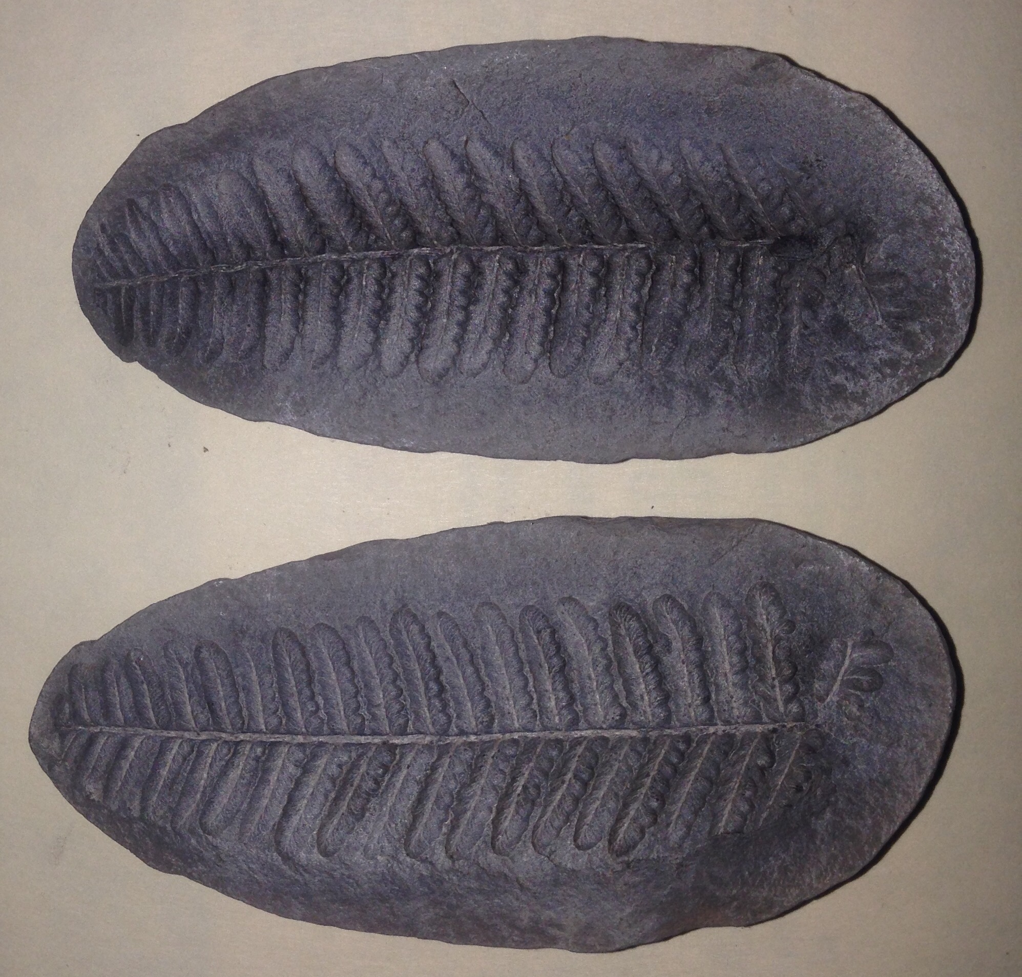 The majority of these plants are from a world famous locality in Illinois called Mazon Creek, which includes sediments from the Late Carboniferous Period (approximately 300 million years old). Through a combination of rapid burial and unusual geochemical conditions organic material at Mazon Creek was exquisitely preserved in hard concretions. The vast majority of the Mazon Creek fossils are plants (and the collections donated to WSC only includes plants) but many animals have also been recovered, including marine invertebrates (even soft-bodied jellyfish), insects and other terrestrial invertebrates, and occasional fish and sharks.It will take us awhile to sort and identify all the specimens in the donation, which included several boxes of material. The WSC previously had almost no Carboniferous fossils in our collection, so this is a very nice addition. I'd like to thank Andrew Moore and Cynthia Fadem of the Earlham College Geology Department for arranging for the donation of this wonderful collection.
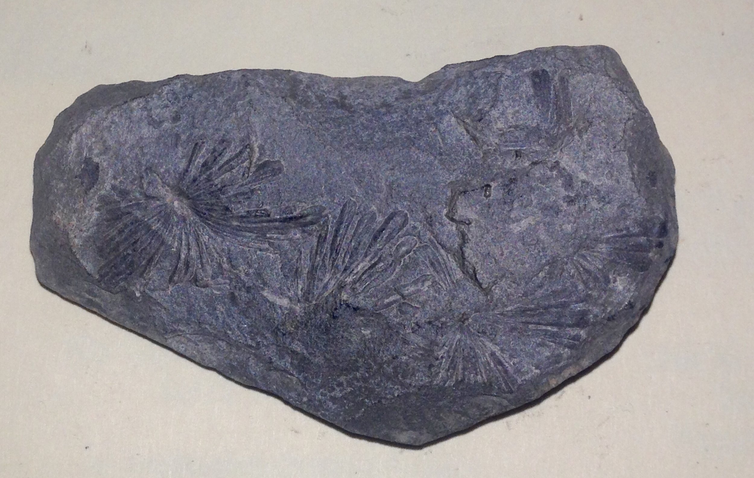
Fossil Friday - rattlesnake vertebra
 It's springtime, and in the Inland Empire that means snakes! The Pleistocene fossils from Southern California make it clear that this has been the case for a long time, as demonstrated by the vertebra shown above.This partial vertebra is probably from the genus Crotalus, a rattlesnake, shownfrom the front (anterior view). Snake vertebrae are procoelous, meaning that the vertebrae articulate with each other with a ball-and-socket joint with the socket on the front of the vertebra; the socket is clearly visible in the image above.Snakes have a lot of vertebrae, up to several hundred in some species, so each individual vertebra is typically not very big. This vertebra is a little over a centimeter across, which represents a pretty large rattlesnake.
SE GSA meeting Day 2
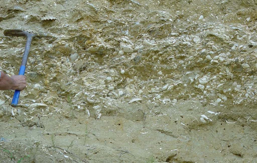 I'm on my way back home from the SE GSA conference, and I finally have a chance to write about the second day of the meeting. Things got very busy at the WSC booth (we sold most of our inventory of casts!), and as a result I missed the entire morning session of talks except for single poster.That poster was by Nickacia Young and Rowan Lockwood on the effects of cementation on the preservation of fossil shells in Late Miocene deposits on the Virginia Coastal Plain. These deposits, called the Eastover Formation, are extremely rich in fossil shells. They are usually unlithified, meaning that the sediments are soft and can be dug out with a pick and shovel, such as in the image above. But in a few places the Eastover is lithified, meaning that it has fused into solid rock, as with the orange blocks in the image below:
 According to Young and Lockwood the number of species of shells (the diversity) in the lithified Eastover is much lower than in unlithified samples. This could have implications in estimating biodiversity in other deposits in which the entire deposit is lithified; if it behaves like the Eastover we may only be seeing a small subset of the species that were originally present.In the afternoon I pick up a couple of talks in a session on faults and shear zones in the Appalachians. John Hickman discussed deeply buried fault zones beneath Kentucky, and suggested that they form part of a Precambrian rift system associated with the breakup of the supercontinent Rodinia in the Late Proterozoic Eon.The next talk was presented by Chuck Bailey, on which I was one of four coauthors. This was on the presence of Mesozoic faults in the Hylas Fault Zone at the edge of the Virginia Coastal Plain and their effect on younger Cenozoic sediments. I was involved in this talk because one of the study sites was the Carmel Church Quarry, a marine fossil site that I have been excavating for more than 20 years. During and excavation I led at Carmel Church in 2014 we discovered a boulder field buried in the Miocene sediments along with whales and other marine fossils:
 I suspected that structural activity might be responsible for the presence of these boulders, and since Chuck was working on faults in this region I asked him to take a look at the boulder field. Based on work by Chuck and his students it appears that the boulder field was most likely formed by the reactivation of a Triassic fault in the Miocene.As soon as Chuck's talk was finished I raced to another lecture hall to catch the end of a session on teaching evolution in the southeast, organized by Patricia Kelley and Christy Visaggi. This was actually an all-day session with 16 talks, and while the booth kept me away from the morning session Brett was able to attend. I had to attend the afternoon session, though, because Brett and I were jointly presenting a talk on teaching activities for getting students used to making hypotheses based on limited data. This included a description of the cast-based teaching kits that Brett and I have been developing based on specimens housed at the Western Science Center, as well as the adaptation of online lessons for courses in historical sciences.The last talks at SE GSA were Friday afternoon. There were also several post-meeting field trips on Saturday, but with long drives ahead of us Brett and I had to start heading back to Virginia and California, respectively. This was a fun and productive meeting, and I have to say that, having attended conferences in dozens of cities, Chattanooga was one of the nicest conference cities I've ever seen.
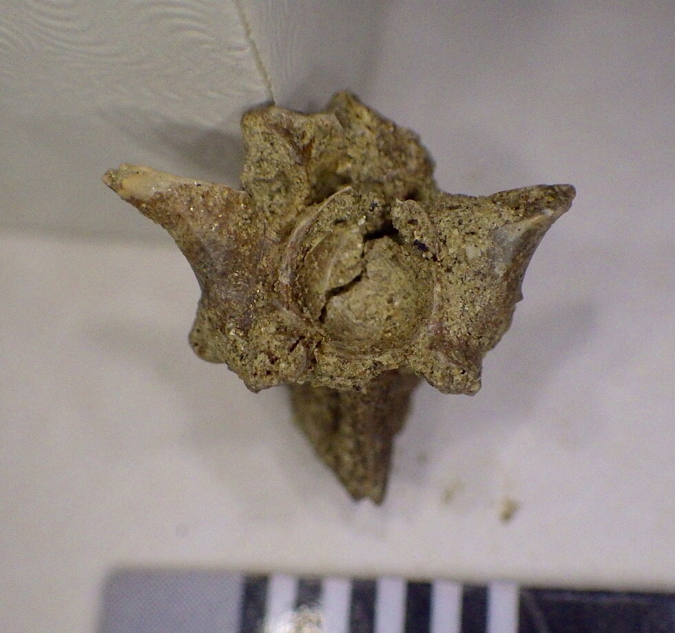
Fossil Friday - nautiloid cephalopod
 Because I'm still at the SE GSA conference today's Fossil Friday post will have to be a short one. Last week Brett and I were in Chicago for the NSTA conference, after which we started driving to Chattanooga for SE GSA. The drive across the intervening states of Indiana, Ohio, and Kentucky took us through some of richest fossil deposits in the world, the Ordovician rocks of the Cincinnati Arch. Practically every exposure of rocks in this region will include at least some marine invertebrate fossils, so even though it was cold and we were pressed for time we still made a quick stop at a road cut in Kentucky. Ten minutes of searching revealed the fossil shown above, a nice natural cast of a nautiloid shell.Nautiloids are cephalopods, the group of mollusks that includes squid and octopus. While most modern cephalopods don't have an external shell, the nautiloids (including the extant chambered nautilus) have a subdivided calcium carbonate shell. While nautiloids are rare today they were much more common and diverse in the past, and during the Ordovician Period were apex predators. But even though they were more common in the Ordovician than they are today, they weren't that common, and usually make up only a tiny percentage of the fossils in most Ordovician deposits. That makes this a very nice find for a single 10-minute stop.