

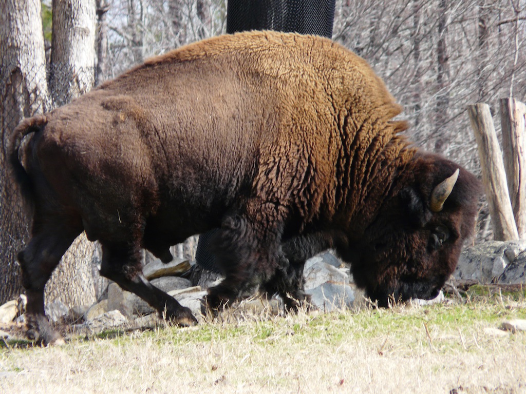
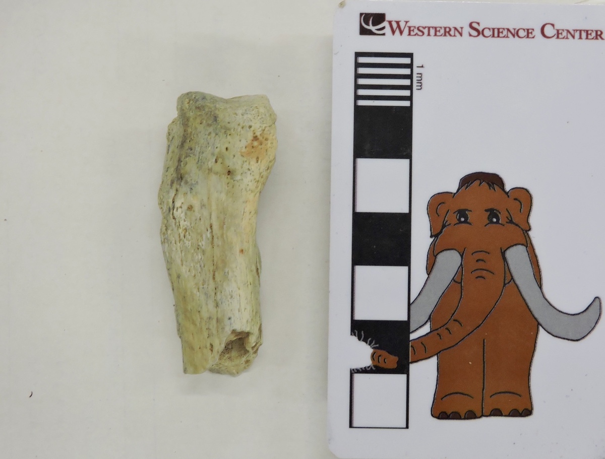
 At first glance, bison seem to be rather oddly proportioned, with a relatively massive head and shoulders. And sometimes, things are exactly as they appear; bison really do have huge, heavy heads. Carrying around such a large head has effects on the rest of the body, and bison have strong neck vertebrae, long neural spines on the vertebrae over the shoulders, and robust forelimbs to help support the weight of the skull. This was even more of an issue for Pleistocene bison such as Bison latifrons and B. antiquus, with their relatively larger and heavier horns compared to the modern B. bison.Today's Fossil Friday specimen is part of the right humerus (upper arm bone) from a Pleistocene bison:
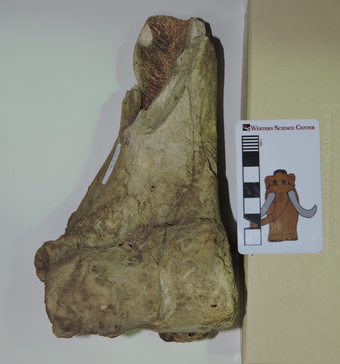 It's shown above from the front (anterior or cranial view). This is only the distal part of the bone, with the elbow joint at the bottom. The proximal half with the ball joint at the shoulder is missing. This is a big, heavy bone; the articulation surface at the elbow is about 9 cm across.Here's the same bone from behind (posterior or caudal view):
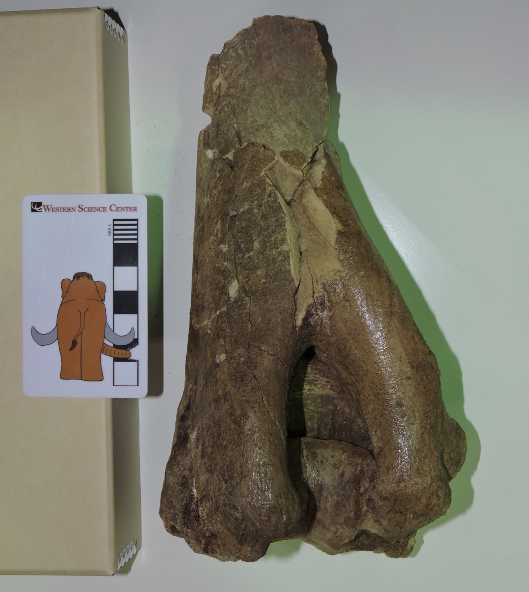 The large notch at the bottom is called the olecranon fossa. It accommodates a large projection on the connecting bone, the ulna, called the olecranon process. This process forms the point of the elbow, and serves as the attachment point for the triceps brachii muscle that extends the forelimb. When the triceps brachii is flexed, it straightens the forelimb, rotating ulna's olecranon process into the humerus' olecranon fossa.
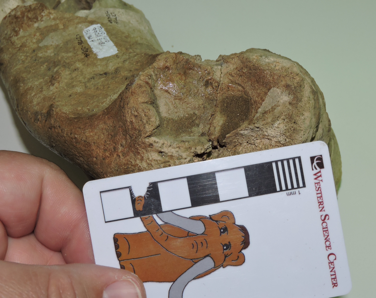 Above is an oblique view of the lateral side of the humerus. The circular area above the scale bar is called the lateral epicondyle, and in this specimen it has a somewhat irregular shape and texture. This could be due to osteoarthritis, which wouldn't be surprising in the elbow joint of a heavy-headed animal such as a bison. The distal end of this humerus is fully fused to the rest of the bone (unlike the mastodon humerus we featured last week), suggesting that this bison was full grown at the time of its death.
Fossil Friday - juvenile mastodon humerus
 It's been over two months since we've featured a mastodon for Fossil Friday, which seems a little odd for the Valley of the Mastodons, so this week we have a mastodon humerus.This is a left humerus (the upper arm bone), seen above in approximately anterior view. The proximal end (closest to the shoulder) is on the left, while the distal end (closest to the elbow) is on the right. Below is the same bone in posterior view:
 The ends of the bone are indistinct and have no obvious articulations with other bones. While there is some damage to the bone, the primary reason for this is that this was a very young mastodon. The humerus starts out as three different bony components, a main shaft and epiphyses at each end that are all held together by cartilage. As the animal grows the elements eventually fuse together into a single unit, but if the animal dies before it's fully grown the epiphyses may fall off as the cartilage decays. That's what's happened in this case, with both the missing epiphyses and the small size indicating that this was a very young mastodon.
Fossil Friday - Smilodon metacarpal
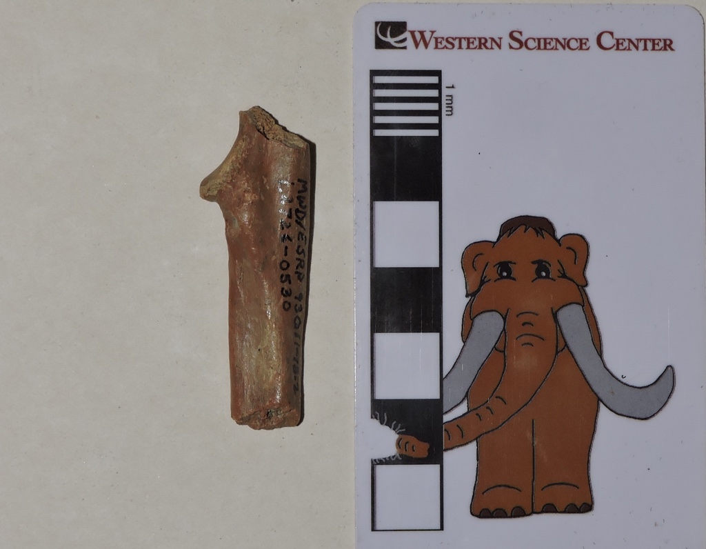
 Part of the nature of paleontology is attempting to pull as much information as possible out of often very limited data. As I mentioned last week, many fossil specimens may not be especially visually compelling, but nevertheless can provide useful information.A few weeks ago I discussed a bighorn sheep bone from a small collection of fossils recovered from Bureau of Land Management property near Desert Center in Riverside County. A number of other species were found at this site, with one of the most surprising being the sabertooth cat Smilodon.The only identifiable fragment from Smilodon that was found was the partial left second metacarpal (the hand bone that supports the index finger) shown above. It happens that at WSC we have a cast skeleton of Smilodon that we use for programming that allows us to place it in context:
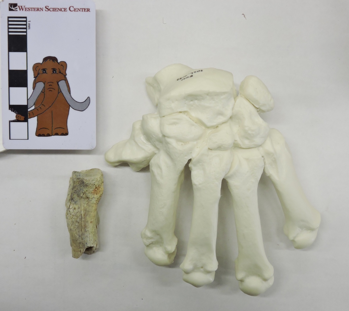 Here's a marked-up version, with the approximate preserved portion of the metacarpal indicated:
 So all we have is the proximal half of the bone, and even that is damaged. Nevertheless, the size and shape make this a pretty solid identification. The interesting thing is that this is the only reported Smilodon bone from the entire region around Desert Center, and in fact is the only record of any large Pleistocene cat from this area (Raum et al. 2014). So this tiny fragment turns out to be a relatively significant specimen for what it can potentially tell us about the range of Smilodon and the Pleistocene ecosystem in the Desert Center region.Reference: Raum, J., Aron, G. L., and Reynolds, R. E., 2014. Vertebrate fossils from Desert Center, Chuckwalla Valley, California. In R. E. Reynolds (Ed.), Not a Drop Left to Drink, California State University Desert Studies Center 2014 Desert Symposium: 68-70.
Fossil Friday - Stories from Bones exhibit
![]() For Fossil Friday this week, I want to highlight Western Science Center's new exhibit "Stories from Bones", which opens tomorrow.While WSC has excellent paleontology exhibits, as with any museum with a large collection many of the specimens are not on public display. There are a variety of reasons for this. Of course, the biggest obstacle is money; cases, information panels, interactive, floor space, and other requirements for an effective display are all expensive, and even the healthiest museums operate on a shoestring budget. Besides money issues, many specimens are just not suitable for display. Perhaps they're too fragile to risk moving around too much, or too fragmentary to interpret for the public (a specimen that visually looks like a piece of junk can still produce valuable scientific data). Even with all these limitations, we strive to make as much of our collections accessible to the public as possible. "Stories from Bones" is a result of that effort.
For Fossil Friday this week, I want to highlight Western Science Center's new exhibit "Stories from Bones", which opens tomorrow.While WSC has excellent paleontology exhibits, as with any museum with a large collection many of the specimens are not on public display. There are a variety of reasons for this. Of course, the biggest obstacle is money; cases, information panels, interactive, floor space, and other requirements for an effective display are all expensive, and even the healthiest museums operate on a shoestring budget. Besides money issues, many specimens are just not suitable for display. Perhaps they're too fragile to risk moving around too much, or too fragmentary to interpret for the public (a specimen that visually looks like a piece of junk can still produce valuable scientific data). Even with all these limitations, we strive to make as much of our collections accessible to the public as possible. "Stories from Bones" is a result of that effort.
 Mammoth jaw display in "Stories from Bones".
An important aspect of planning an effective exhibit is developing a theme. An exhibit is telling a story, and you need to be aware of what that story is as the exhibit is being designed. The theme might be "We have a bunch of stuff!", but while that was a common theme in museums a century ago (and one I personally appreciate), it does not generally make for the most informative exhibit experience for the majority of visitors.Once the theme is established, it's important to stick to it, so that the exhibit story remains coherent. Imagine reading a mystery novel in which three chapters are devoted to a history of the development of the gunpowder used in the crime, and an additional chapter describes the etymology of the last name of the victim, when neither is important to the outcome of the story. Each of these things might be individually interesting, but if you try to talk about all of them then you risk obscuring everything. There is a real risk of this "mission creep" in an exhibit based on a data-rich field such as paleontology. We might talk about evolutionary relationships, paleoenvironmental indicators, biogeographic information, site-specific descriptions, or an array of other things. Talking about any of these might be a good idea; talking about all of them is a bad idea.The permanent paleontology exhibit at WSC does this very well. The exhibit is basically a review of the Diamond Valley Lake Local Fauna; what was here, how does it compare to the rest of Southern California, and (as a secondary point) what does it tell us about the local Pleistocene paleoenvironment. In contrast, "Stories from Bones" asks "What do these fossils tell us about the lives and deaths of these individual animals?".To that end, "Stories" has a series of displays that talk about how paleontologists determine how old an animal was when it died. We have several cases that look at tooth replacement in proboscideans, horses, and bison, such as the two mammoth jaws above (they're close to the same size, but one animal was about 30 years older than the other), or the three bison dentaries shown below that represent young, middle-aged, and elderly animals.
 Bison jaw display in "Stories from Bones".
We have several examples of bones that were broken and healed, evidence of events that took place during an animal's life:
 Broken and healed bones in "Stories from Bones".
We also have several cases that describe taphonomic features, looking at what happened to an animal at or immediately after death.We designed and built a number of interactive displays for this exhibit. The most prominent is a cast and video of the CT scans of Max the Mastodon's lower jaw, taken back in August.
 Max's CT-scan station in "Stories from Bones" during installation, under the watchful eye of @MaxMastodon.
We're proud of the fact that several of our interactive displays ask visitors to map or measure specimens and reach conclusions based on their data:
 A more extensive version of the bison tooth display shown here is also available as a guided activity for school groups visiting the museum, and as a kit available for purchase.If you're a regular reader of this blog, you'll find that "Stories from Bones" draws heavily from my past "Fossil Friday" posts. For most of those specimens, this is the first time they've ever been on public display, so if you're near Southern California make sure to stop by the museum. "Stories from Bones" opens on October 31, and will remain open into May 2016.
Fossil Friday - horse deciduous premolar
 Last summer I posted about a partial horse skull from Diamond Valley Lake that still had its deciduous premolars in place. Of course, if that horse had lived a little longer the deciduous teeth would have fallen out and been replaced with permanent teeth, with the remnant of the shed tooth left behind.The large tooth shown above is a shed deciduous premolar, probably the upper left dP4. Besides Max, for scale there is also a modern human deciduous premolar (P2 instead of P4, since humans only have 2 premolars) courtesy of my son Tim.Horses eat abrasive food such as grasses, and their teeth take a beating. By the time the 4th deciduous premolar is shed at around 3 years of age, it is worn to a nub. This is more apparent in an oblique view of the same tooth:
  In the Diamond Valley Lake collections we only have a few shed deciduous horse teeth. Many animals don't live enough to shed their teeth, and if they do live that long, by the time the teeth are shed they're worn so thin that they aren't as likely to survive to become fossils as a more complete tooth.This tooth will be on display in the "Stories from Bones" exhibit, opening at Western Science Center on October 31.
Fossil Friday - Alamosaurus neck vertebrae
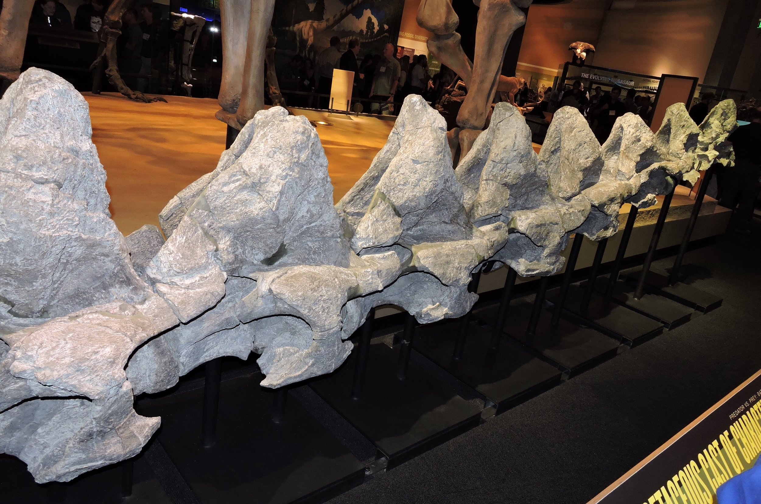 This week I've been attending the annual Society of Vertebrate Paleontology (SVP) meeting, which is being held this year in Dallas. SVP usually is held in a city with a natural history museum, and on Wednesday night the local museum will generally host a welcome reception. So for this week's Fossil Friday I photographed one of the highlights of the Perot Museum of Nature and Science's exhibits, the neck of Alamosaurus.Alamosaurus was a large sauropod dinosaur that has been found in Cretaceous sediments in the southwestern United States; this specimen is from Big Bend National Park in Texas. While not as famous as its older Jurassic relatives like Brontosaurus, Alamosaurus was just as large and impressive. This is easy to see when you realize that in the photo above you can only see 8 neck vertebrae!This image is taken as if you're standing near the base of the neck on the right side, looking forward, so the head would be toward the far right. There were at least a few more vertebrae at the base of the skull that were not preserved in this specimen, and more at the base of the neck as well. In fact, there is a ninth vertebra on exhibit, but the neck is so large that with the reception crowds I couldn't get far enough away to get all nine vertebrae in one photo from this angle. So here are all nine, looking from the front to the back:
  The Perot Museum asked Research Casting International to produce a cast reconstruction of Alamosaurus based on these vertebrae and other Alamosaurus specimens. The cast is also on exhibit:
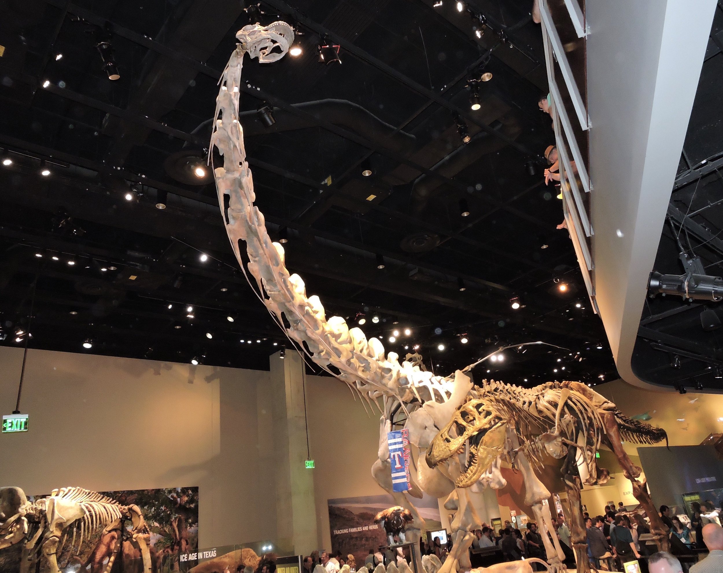 The people standing around the exhibit give an idea of scale. If that's not enough, that little theropod dinosaur standing beside Alamosaurus is Tyrannosaurus, and a Columbian mammoth is just visible at the lower left. By any standards, Alamosaurus was a huge animal!For more abut Alamosaurus, and to see photos of this specimen in the field, visit the SV-POW! blog.
Fossil Friday - badger ulna
 It happens that last Tuesday, October 6, was National Badger Day in Britain. While the European badger Meles meles does not occur in North America, why should that stop us from honoring badgers? The American badger Taxidea taxus (above at The Living Desert Zoo) is only distantly related to the European badger, but it still goes by the common name of "badger", and that's good enough for me!Taxidea is widespread in North America today, so it's no surprise that is occasionally turns up in Pleistocene deposits. A handful of badger bones were found in the Diamond Valley Lake excavations, including this right ulna:
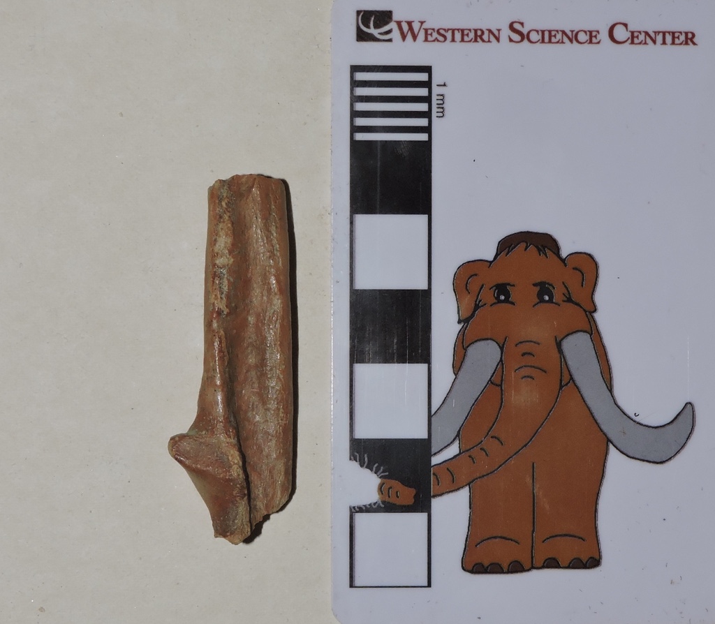
 This fragment represents roughly the middle third of the ulna, one of the bones in the forearm. If that doesn't seem like much, keep in mind that badger bones make up roughly 0.0015% of the bones collected from the Diamond Valley Lake deposits, so even with such a small piece we're doing pretty well!
Fossil Friday - deer metacarpal
 One point I often come back to is how the Pleistocene fauna is in many ways so similar to today's fauna. Geologically speaking, the Ice Age is the recent past; many of the species alive then are still with us today.The bone shown above is the right metacarpal (forefoot bone) of a deer from the genus Odocoileus. Odocoileus is the most common large wild mammal in North America today, with the white-tailed deer O. virginianus found through most of the continent and into South America, and the mule deer O. hemionus ranging throughout western North America. While mule deer tend to be a bit larger than white-tailed deer (at least today), it's difficult if not impossible to distinguish between them based on limited remains such as this.This metacarpal is incomplete, with the proximal portion (closest to the wrist) broken off. The image above is in anterior view, while below is the posterior view:
 At the distal end of the bone, note that there are two articulations. Deer are members of the Family Cervidae, which are in the Order Artiodactyla. Like their relatives the Bovidae (cattle and their kin), Camelidae (camels), and many other artiodactyl groups, deer have two digits on each foot, digits III and IV (the middle and ring fingers on the hand, and the equivalent toes on the foot).Deer were not a major component of the fossil fauna at Diamond Valley Lake; horses, bison, camels and even mastodons are more common as fossils in these deposits. Nevertheless, they were common enough for multiple specimens to show up in the sample.
Fossil Friday - bighorn sheep toe bone
 Earlier this week Western Science Center received a small collection of fossils recovered by Paleo Solutions from Bureau of Land Management property near Desert Center in eastern Riverside County. While the material was limited, some was identifiable, including a few unusual pieces.This particular specimen is the first phalanx (toe bone) from what appears to be a bighorn sheep, Ovis canadensis, identified as such in Raum et al., 2014 (modern example below from a herd at Badlands National Park):
 Historically bighorn sheep were widespread in the mountains and deserts of the American west, although their populations are now greatly reduced. There are still several hundred living in Joshua Tree National Park, not far from where this fossil was found.Fossil bighorn sheep do not seem to be particularly common; this is the only one in the WSC collection (as far as we know). This specimen is most likely Pleistocene (rather than something more recent) because extinct Pleistocene animals such as Smilodon were found in the same deposit.Reference: Raum, J., Aron, G. L., and Reynolds, R. E., 2014. Vertebrate fossils from Desert Center, Chuckwalla Valley, California. In R. E. Reynolds (Ed.), Not a Drop Left to Drink, California State University Desert Studies Center 2014 Desert Symposium: 68-70.
Fossil Friday - horse metatarsal
 While the bulk of the Western Science Center's paleontology collection comes from Diamond Valley Lake, we have significant collections from other localities. One of the most interesting collections comes from Southern California Edison's El Casco Substation in northern Riverside County. This material, while probably still Pleistocene, is well over a million years old, about 4 to 6 times older than anything recovered from Diamond Valley Lake.Horses are among the most common remains from El Casco. Shown at the top is a horse left metatarsal (foot bone), seen in anterior view. The bone is lying on its side, with the proximal end (closest to the ankle) on the right and the distal end (closest to the toe) on the left. The bone is somewhat deformed, so even though this is anterior view, part of the left side is visible (the edge closest to the scale bar). Notice that the bone looks a bit swollen at one point, adjacent to where Max the Mastodon is holding the scale bar.Here's the same bone in posterior view:
 Again, the proximal end of the bone is on the right. In this view we can see that three bones are actually present, one large bone in the middle and a slender bone (commonly called "splint bones") on each side. In fact, all three of these bones are metatarsals.All modern horses in the genus Equus (and fossil Equus like this one) have a single functional toe on each foot. Like all mammals, horse ancestors had five digits on each hand and foot, but over time most of those digits have been lost. The one remaining functional digit is the third one, the middle finger on the hand and middle toe on the foot. These are attached to the 3rd metacarpal (hand) and 3rd metatarsal (foot).But what about those splint bones? Well, horses may only have one functional toe on each foot, but they still have remnants of the second and fourth metacarpals and metatarsals, which have not yet been completely lost. In the bone shown above, from the top you can see the 4th metatarsal, the 3rd metatarsal (by far the largest), and the 2nd metatarsal. The swollen area that is so visible in the top photo occurs near the distal tip of the 4th metatarsal, more clearly visible in lateral view:
 The distal tip of the 4th metatarsal is fused onto the side of the 3rd metatarsal. This is a pathological condition, is is likely a healed fracture of one or both of these bones. In medial view the 2nd metatarsal shows the normal condition, where it lies close against the 3rd metatarsal but with no fusion at the distal end:
 This injury was clearly not fatal, since it healed. There's no telling what caused the injury, but some type of blow to the outside of the foot is possible; maybe it was kicked or stepped on by another horse. But it does give us a snapshot, if an incomplete one, into the life of this horse.This specimen, along with many others in our collection, will be on display in our new exhibit Stories from Bones, which opens at the Western Science Center on October 31.
Fossil Friday - mammoth molar
 After a few weeks of focusing on mastodon remains, it's worth remembering that there were two different species of proboscideans at Diamond Valley Lake. While mastodon remains are quite common, Columbian mammoths (Mammuthus columbi) were also present.This specimen is a lower left 1st or 2nd molar of M. columbi, discovered during the early years of the DVL excavations. Like the closely related Asian elephant, mammoths had enamel that is folded multiple times, wearing to produce a series of raised transverse enamel ridges in occlusal view. Like other advanced proboscideans mammoths replaced their teeth sequentially, so the front of the tooth (on the right in the image above) is more heavily worn that the back of the tooth. In fact, the last enamel ridge is essentially unworn, so this tooth had probably not quite completely erupted.A cast of this tooth is on permanent exhibit at the Western Science Center, as a touchable display allowing visitors to compare the very different occlusal surfaces of mammoth and mastodon teeth. As it happens, we're also auctioning off an additional cast of this tooth (below) tomorrow night at our annual Science Under the Stars fundraiser.
Fossil Friday - Max's mandible, CT-edition
 Last week we took Max to California Imaging and Diagnostics in Hemet, who donated a some time on their CT scanner to scan the lower jaw of Max the mastodon. A couple of days later CID provided us with disks with the scan data, and we've finally had a chance to start examining them. If you're unfamiliar with CT scans, the term is short for X-ray computed tomography. CT scans take a large number of X-ray images from multiple angles. A computer can then combine those images to make cross-section X-ray slices through the object. The slices can be stacked to make a digital 3D model of the object, or removed to examine the internal structure at any particular slice. It's an invaluable, non-destructive method of examining the inside of an object.I've uploaded an initial video of the slices of Max's jaw. The slices are transverse, run anterior to posterior, and are viewed facing anteriorly. Basically, imagine standing behind the jaw looking forward, and seeing cross-sections of the jaw gradually getting closer to you:http://www.youtube.com/watch?v=hK8c5pAgUrsSince the video runs front to back, the first thing you see is the anterior tip of the jaw. (Well, actually the the first thing is the plaster cradle the jaw is sitting on, then the tip of the jaw.) As you move back, you pass though the mandibular symphysis where the two haves of the jaw are joined, through the teeth, and then to the coronoid process and mandibular condyles (the condyles are incompletely shown, because the jaw was a bit to large for the scanning area).There are several slices that have especially noteworthy features. This is about 4 seconds into the video:
 The image of the jaw on the right is for orientation; the purple line is the approximate location of the slice. The blue arrow is pointing the pathological bone growth near the tip of the right dentary. Inside the bone, there are a series of fractures next to this bony growth, one of which is marked with the green arrow. I'm not sure if they are actually associated with the growth, or if they occurred after burial.Moving a little further back, to about 6 seconds:
 We're now behind the bony growth, but its effects are still present. The blue arrow is pointing to a foramen (a passage for nerves or blood vessels) that has been displaced anteriorly and ventrally, apparently in response to the growth. The green arrow is pointing to the mandibular symphysis, where the left and right dentaries are joined together. This was a surprise to me. These bones fuse together in older animals, obscuring the line of fusion. Max was a fully mature mastodon, middle aged at least. I would expect the symphysis to be completely fused, and on the outside it is; there's no trace of the symphysis on the outside of the bone. Yet inside they're still unfused. Is this typical of mastodons, or the Max's symphysis never fuse (or pop open) due to the beating his jaw apparently went through?Moving further back, to about 8 seconds:
 This slice shows how much that foramen was displaced, when compared to the left dentary (blue arrows). It's also clear that the dentary is still messed up well behind the bony growth; note how asymmetrical the two dentaries are on the dorsal surface (they should be nearly mirror images). In fact, the entire mandible is quite asymmetrical. I originally assumed that this was mostly due to deformation that occurred after burial, but it's now clear that at least some (maybe most) of the asymmetry occurred while Max was alive.At around 14 seconds:
 We're now getting into the tooth rows. The first root and cusp of the left 2nd molar is clearly visible (yellow arrow). The white arrow below the tooth is the opening of another foramen. One the right, the notch marked by the green arrow is the socket for the last root of the right 1st molar. Max was a mature mastodon, so the 1st molars (and all the premolars) had already worn down and fallen out; only the 2nd and 3rd molars remained, and the 2nd molars were heavily worn.As we already knew from examination of the exterior, Max had a second injury on his right dentary, essentially a crease running obliquely below the anterior tooth row with ridges of secondary bone growth on each side of the crease. This injury is marked with the blue arrow, showing apparently compressed bone beneath the surface (the bright line on the inside) and the swelling associated with the secondary bone growth.From this point in the video you can see the tooth cusps and roots grow and shrink as the scans run through the tooth rows. There is a change in one of the teeth at around 28 seconds:
 At this point we're all the way back to the last root of the 3rd left molar. As indicated by the green arrow, the pulp cavity on this tooth is open (you can see the same thing on the last root on the right 3rd molar at 29 seconds). This suggests that this part of the tooth was still growing. Sure enough, if we look at the cusps, the last cusp on the 3rd molar is only very slightly worn, indicating that it had just erupted.By 34 seconds the scans have passed completely through the tooth rows, and the remainder of the scans pass through the coronoid processes and the mandibular condyles.We still have a lot to look at with these scans, and we're hoping they'll be useful to people that are working on mastodon anatomy. We're going to keep working to make this data available online as we have time to process it. We also intend to include annotated versions of these scans in our upcoming exhibit, Stories from Bones.
Fossil Friday - Max the mastodon's mandible
 If you follow the museum's social media pages, you' re probably noticed that last Tuesday we had an adventure with one of the museum's mastodons, Max. We pulled Max's lower jaw off exhibit and, via ambulance and with a police escort, sent him to California Imaging and Diagnostics in Hemet for X-rays and CT-scans. There were several reasons behind this effort, but the primary one was that Max's jaw shows evidence of several injuries that we wanted to further explore.
 Max leaves the museum amid a crowd of Western Center Academy students.
 Preparing to load Max into the ambulance. One of Max's injuries is very obvious and we've known about it for some time. There is an anomalous bony growth at the right anterior tip of the jaw:
 The ridge anterior to the teeth on the dorsal side of the jaw is also rather misshapen in this area, possibly related to the same condition.The second injury was first spotted a few months ago, and was actually first observed in a cast we were making of the jaw; in the original specimen the injury was hidden due to the case structure and lighting the exhibit. This injury is a 2 cm-wide groove, running diagonally along the right dentary below the second molar. There are ridges of swollen secondary bone (a healing response to a bone injury) on each side of the groove:
 We're still processing the data gathered at CID, and I'll have more on that (including images) in a future post. But there are already some things we can say about these features, starting with the fact that they occurred while Max was still alive, at least weeks or months before his death(and possibly years before). That's because both feature show evidence of bony growth, and it takes time for that to occur. Evidence of healing is the most reliable method of determining whether a break in a fossil bone occurred before or after death.Max is a large mastodon, the largest reported from California. Given the size and the massive tusks it's probably that Max was a male mastodon. That, and the fact that he was a mature adult (based on tooth wear), suggests a possible cause of these injuries: intraspecies combat. Like modern elephants, there isabundant evidence that male mastodons engaged in intraspecies combat (see Fisher, 2009 for example), often resulting in injuries, sometimes fatal injuries. It's possible that both of these injuries, especially the groove on the side of the jaw, were caused by the tusks of another mastodon.Reference:Fisher, D. C. 2009. Paleobiology and extinction of proboscideans in the Great Lakes Region of North America. In G. Hayes (ed.), American Megafaunal Extinctions at the End of the Pleistocene, Springer Science: 55-75.
Fossil Friday - Xena the mammoth's tusks
 "Xena" is the most complete Columbian mammoth specimen recovered from Diamond Valley Lake, including a nearly perfect skull and lots of postcranial material. Both of Xena's tusks were also recovered, and are on display with the rest of her skeleton at the Western Science Center.Elephants and their relatives (proboscideans) use their tusks in all kinds of ways, including fighting, stripping vegetation, and digging water holes. While the tusks are modified incisor teeth, they do not have enamel (some extinct lineages did, but that's another story), so tusks are typically heavily worn at the tips. The polished wear facets are easily visible in the image above.Another curious feature of proboscideans is that, in most individuals, one of the tusks tends to be more heavily worn that the other. In our mounted cast of Xena, you can see that her left tusk is more heavily worn that the right one, as it's blunter and somewhat shorter:
 Since proboscidean tusks are worn during behavioral activities, it's possible that the differential tusk wear is evidence for lateralization, or handedness. Most animals show a preference for using one side of their body over the other. This can be expressed in a variety of ways, including a preference for using a particular hand for detailed tasks in humans, or for using one side of the mouth in feeding in whales. For some unknown reason, in almost all species that show lateralization, right-handedness is more common. Modern elephants are also lateralized; an elephant will always tend to curl its trunk in the same direction when pulling up grass, for example. Differential tusk wear suggests that they also show handedness in behaviors that involve the tusks. In Xena's case, the more heavily worn left tusk suggests that she was left-handed.We recently produced a cast of Xena's left tusk, which will be auctioned off at the Western Science Center's annual Science Under the Stars fundraiser on September 12. If your in Southern California consider coming the the event to support the museum!
Fossil Friday - coyote humerus
 As I've mentioned in several other posts, fossil carnivores are rare in the Diamond Valley Lake fauna, but they're not completely absent. The remains tend to be isolated bone fragments, but they do reveal the presence of the particular taxon. The small fragment shown here is part of a right humerus from a canid (the dog family). Both ends of the bone are missing, but this fragment comes from closer to the distal end (towards the elbow). Three weeks ago I posted about another canid right humerus, probably from a dire wolf. It's useful to see these specimens side-by-side:
 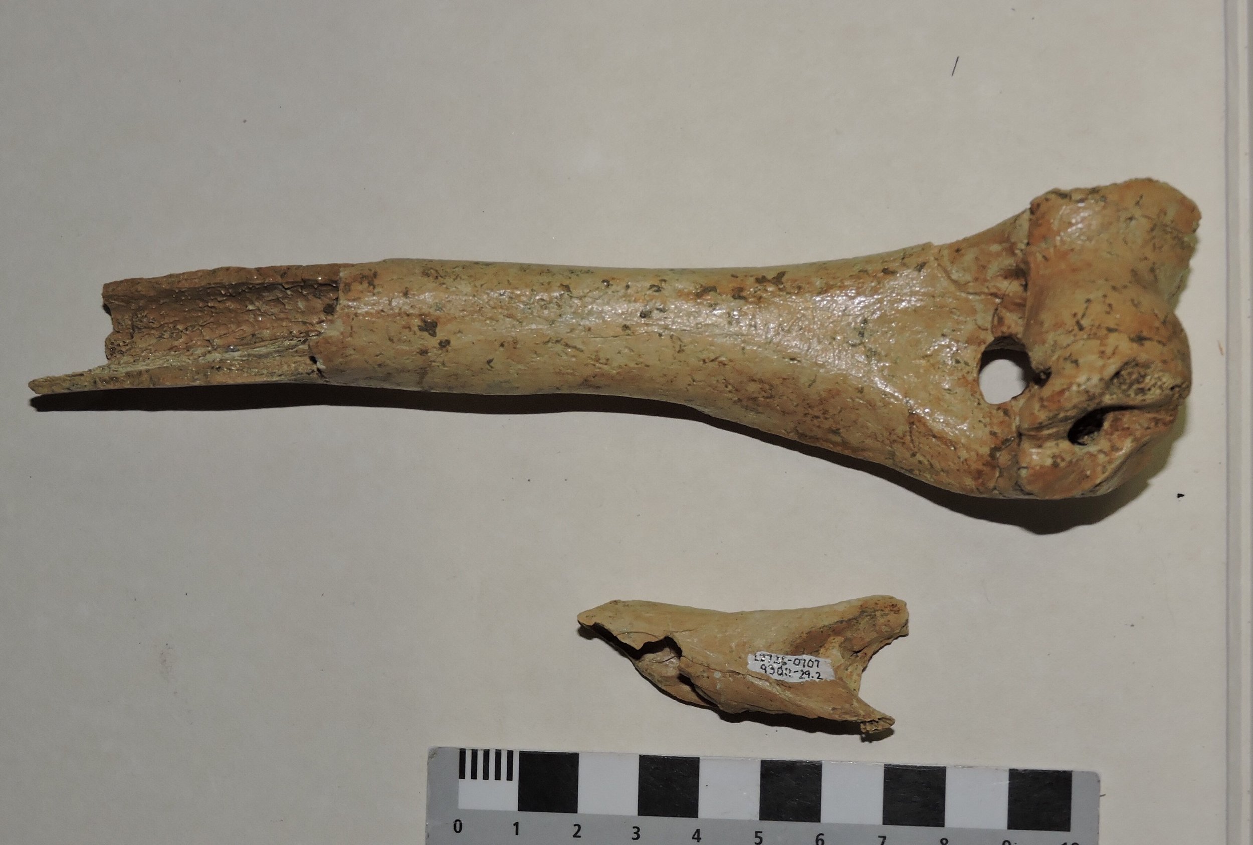 Even though this week's specimen is fragmentary, it's clearly smaller than the dire wolf. Its size is consistent with Canis latrans, the coyote. Coyotes are among the most common mammals at Rancho la Brea, and are still common in Southern California, so their presence in the Diamond Valley Lake fauna is not surprising. But it's still nice to confirm their presence, especially as modern coyotes seem to fill interesting roles in modern ecosystems. There are strong indications that modern coyotes are major predators of pronghorn, but only in the absence of wolves. Apparently wolves alter the behavior of coyotes (either by attacking them or driving them away), resulting in less overall pronghorn predation. In turn, it seems that coyotes suppress populations of domestic and feral cats, reducing predation on birds. These studies hint at the complexity of the interactions within ecosystems. With the amazing range of animals in the Diamond Valley Lake fauna there must have been some fascinating biotic interactions.
Fossil Friday - Equisetum
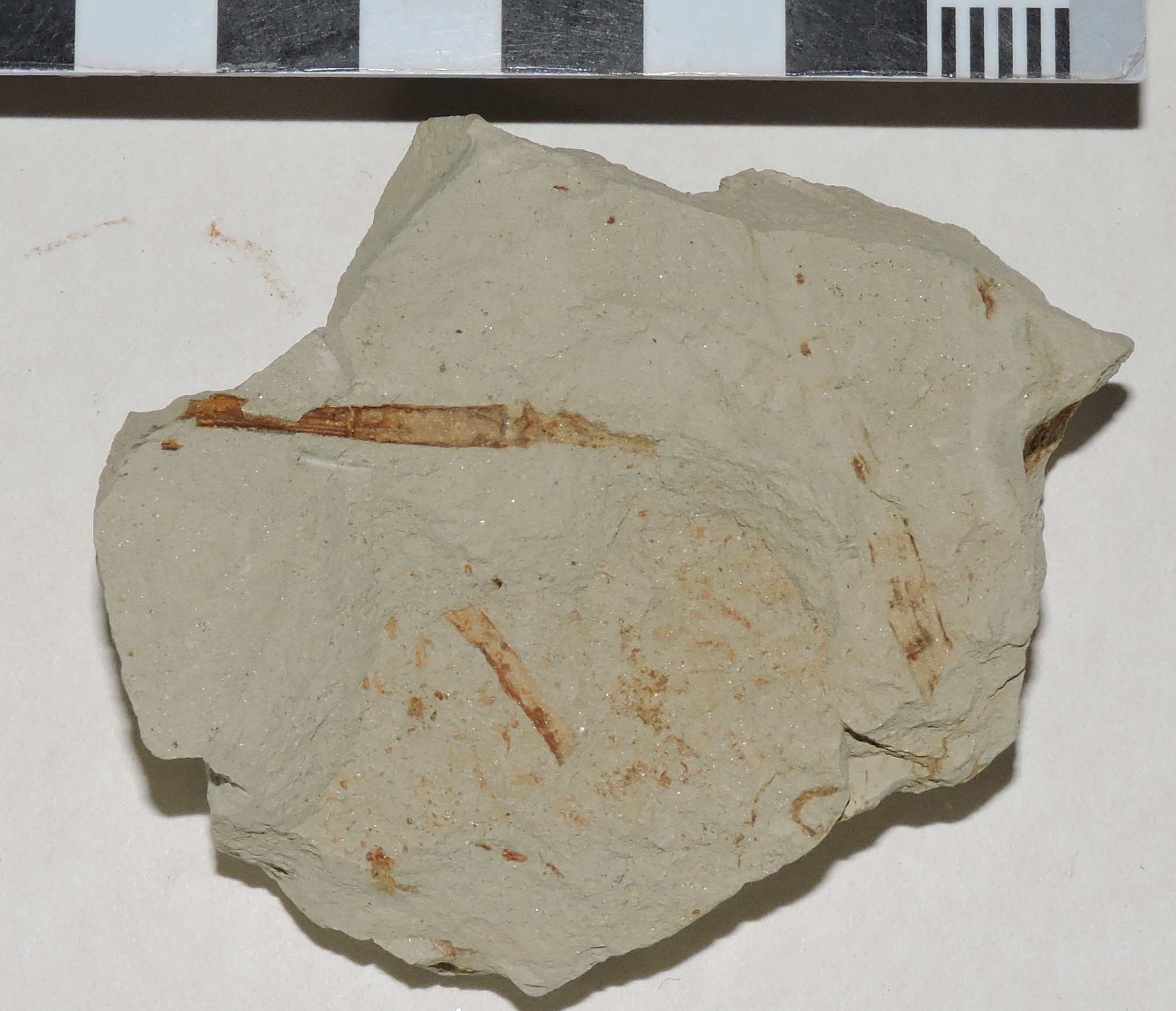 Fossil plants never seem to get the attention they deserve. Fossil animals, especially vertebrates, certainly are fascinating and have tons of things to tell us (they are, after all, what I've worked on for most of my career). But plants are exceptionally good indicators of past environmental conditions, besides being interesting organisms in their own right.Shown above is a sample of the San Timoteo Formation from northern Riverside County, specifically from the Edison El Casco Substation. This excavation yielded several thousand fossils, including plants, animals, and traces, which lived about 1.5 to 2.0 million years ago. The dark brown streaks in this sample are fragments of plant stems. While these don't look like much, there's enough here to make an identification.In the prominent stem toward the upper left, notice the fine grooves and ridges that run lengthwise along the stem (marked with the blue arrow, below). The stem also is divided into segments, marked below with the red arrows:
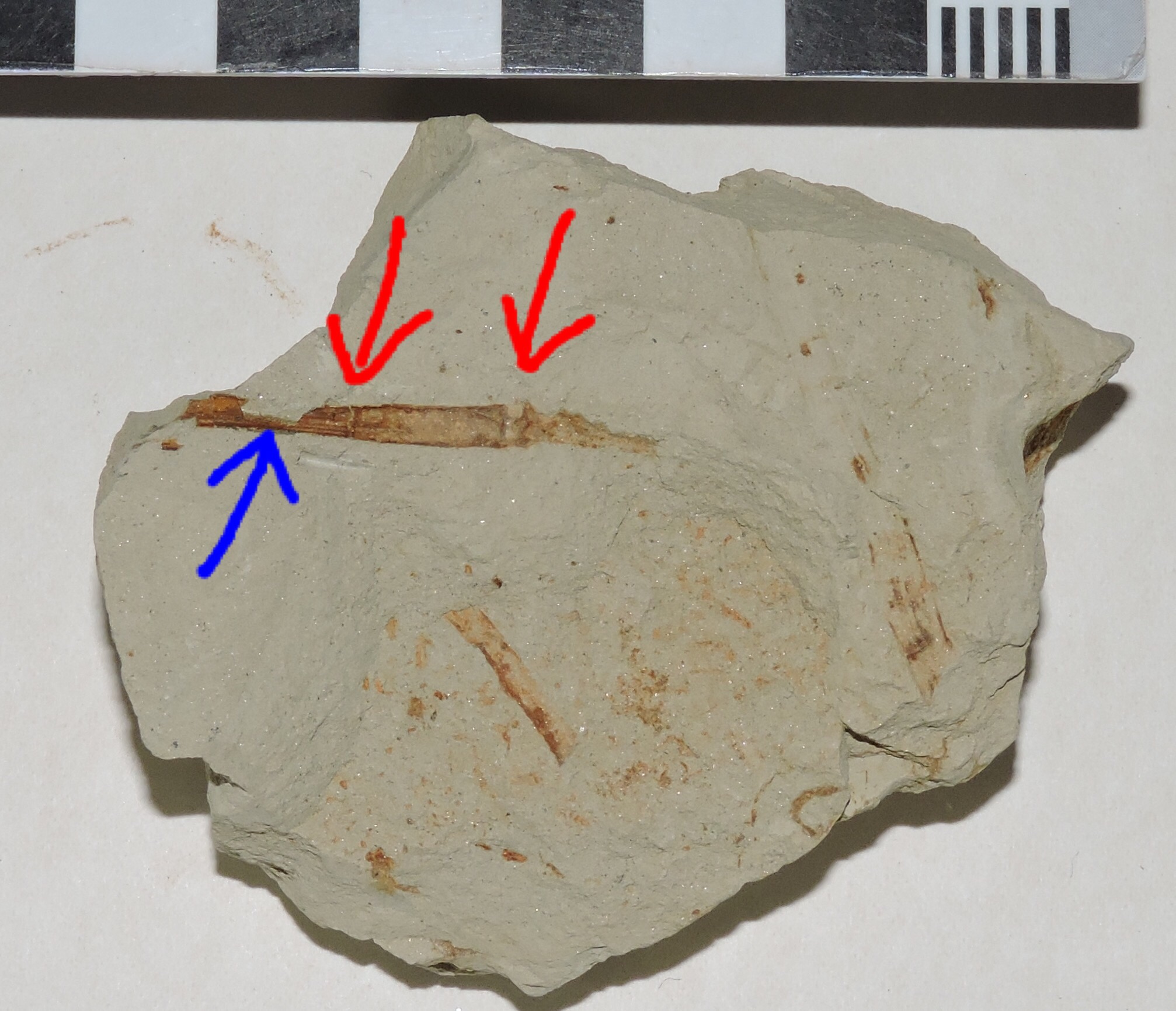 These are both characteristics of the genus Equisetum, commonly known as the horsetail or scouring rush (the latter name comes from the high silica content, which makes them useful as an abrasive). Equisetum is an interesting plant, the last survivor of a group that goes back more than 350 million years. Rather than seeds they reproduce using spores, which are released from a cone-like structure (the sporangium) at the top of the plant. (There may be an impression of a sporangium attached to the stem in this specimen, but I'm uncertain about that.) Horsetails seem to be related to ferns, and according to some genetic studies may actually be highly specialized ferns. Below are some modern examples of the whole plant (at least the above-ground portions), one of which has grown its sporangium. Notice the segmented, striated stems, just as in our fossil example:
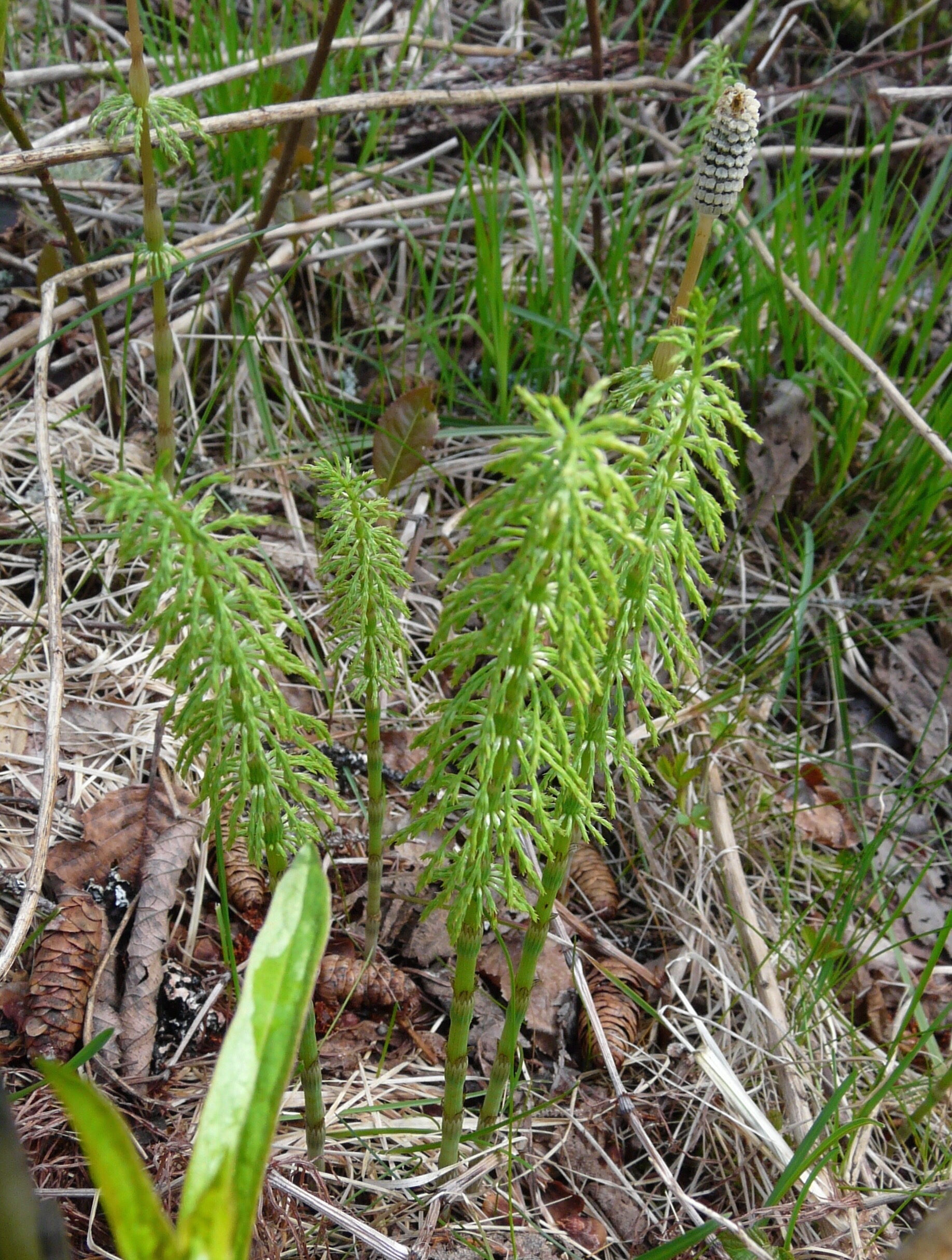 Equisetum also has interesting things to tell us about its ecosystem. Here's a group of modern Equisetum in their typical natural habitat:
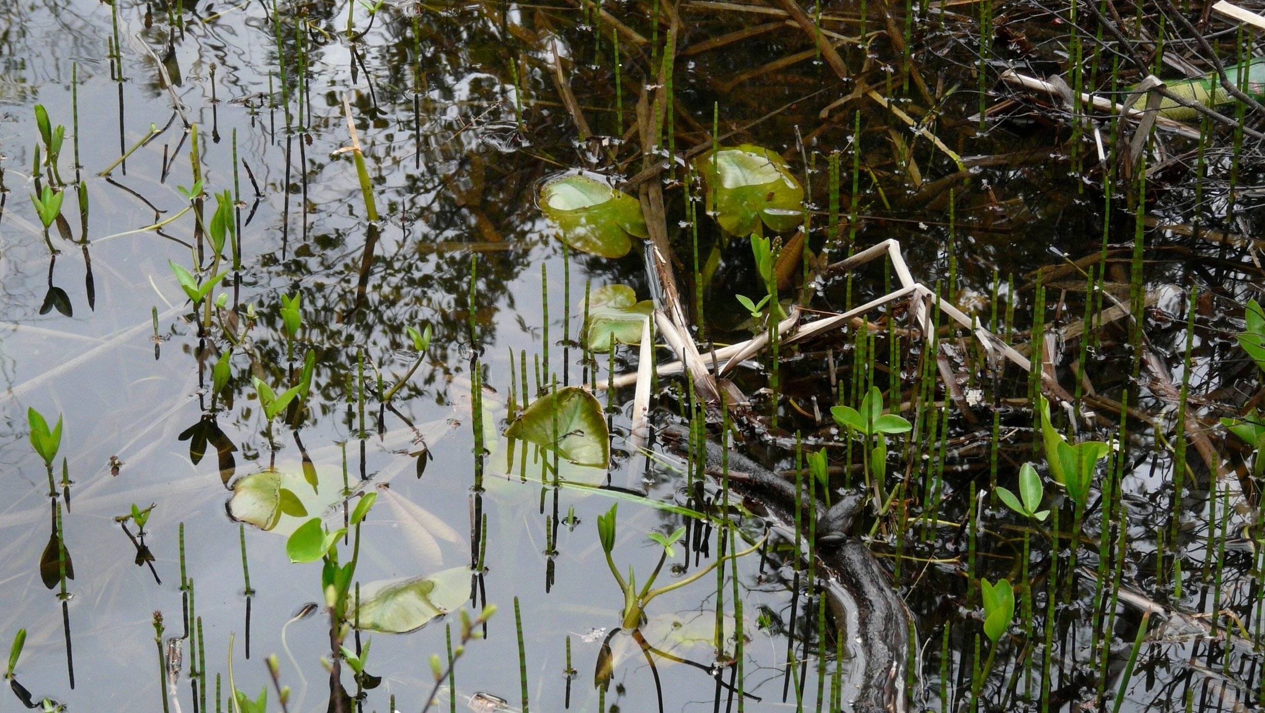 Equisetum loves water, and typically grows in or near ponds or marshes. It seems to be one of the more common plants in the El Casco Substation deposits, and is found with a number of other plant species that prefer similar conditions. This all suggests that, at least locally, conditions there were much wetter than they are today.
Fossil Friday - mastodon caudal vertebra
 Even in huge animals, not every bone is large. This small bone, less than 4 cm in length, comes from one of the largest Pleistocene animals at Diamond Valley Lake — a mastodon.This particular specimen is a caudal, or tail, vertebra. Its simple shape is not due to broken and missing pieces; in fact the bone is almost complete. The prominent spines and processes found sticking out on most vertebrae tend to be reduced or absent on caudal vertebrae in most animals. This is unsurprising from a functional standpoint. Those projecting spines are not simply decoration; they provide anchor points for muscles that control movement of the head, the legs, or other parts of the body. For the most part the tail doesn't serve as the anchor point for any muscles, and so the vertebrae don't generally have large processes. (There are a few exceptions: the first few caudal vertebrae have large processes that anchor the muscles that control the movement of the tail itself. There are also some animals such as alligators that have flattened tails, generally for swimming, which have enlarged caudal spines.) Their simple shape can sometimes make caudal vertebrae tricky to identify. They are sometimes mistaken for toe bones, but in most terrestrial animals even toe bones will have complex articulations and muscle attachment points that caudal vertebrae generally lack.The complete lack of processes indicates that this vertebra is from fairly close to the tip of the tail. To be that far back and still have a length of almost 4 cm indicates that it actually came from a big animal, and in fact other vertebrae associated with this bone indicate that it came from a mastodon.Looking at the bone end-on (below) we can see that the end is fairly rough:
 This is because the bone is missing the vertebral epiphyses, the caps of bone that fit onto each end of the vertebra. The epiphyses remain essentially as separate bones attached to the vertebra with cartilage while the animal is still growing, but eventually fuse onto the vertebra. The fusion of all the various epiphyses indicates that the animal has reached physical maturity; in elephants (and people) this is generally sometime around age 20-25. The lack of fused epiphyses in this vertebra (and, in fact, none of the epiphyses are fused and the other bones associated with this specimen) indicates that it came from a young animal, likely not more than a few years old.
Fossil Friday - canid humerus
 While over 200,000 fossils were found at Diamond Valley Lake, only a tiny percentage of the specimens represent carnivores. This is actually to be expected; in any stable ecosystem prey animals always greatly outnumber their predators and so normally prey animals should be much more common as fossils. There are exceptions, such as "predator traps" like we see at Rancho la Brea, but in general the rare predator/common prey breakdown at Diamond Valley Lake is what we expect to find.That said, there is actually a fairly diverse range of predators from Diamond Valley Lake, even if most of them are known only from isolated bones like as the one shown above.This is the right humerus (upper arm bone) from a canid, the dog family. It's shown above in anterior view, with the proximal end (towards the shoulder) on the left. The proximal half of the bone is actually missing, so we only have the distal half, including the elbow joint at the right end (the condyle). The round hole through the middle of the bone near the condyle is called the supratrochlear foramen. This is a common feature in dogs, but is rare or absent in most other groups, making it a useful identification feature.Below is the medial side of the same bone, showing the sinusoidal shape that is typical of the humerus in carnivores:
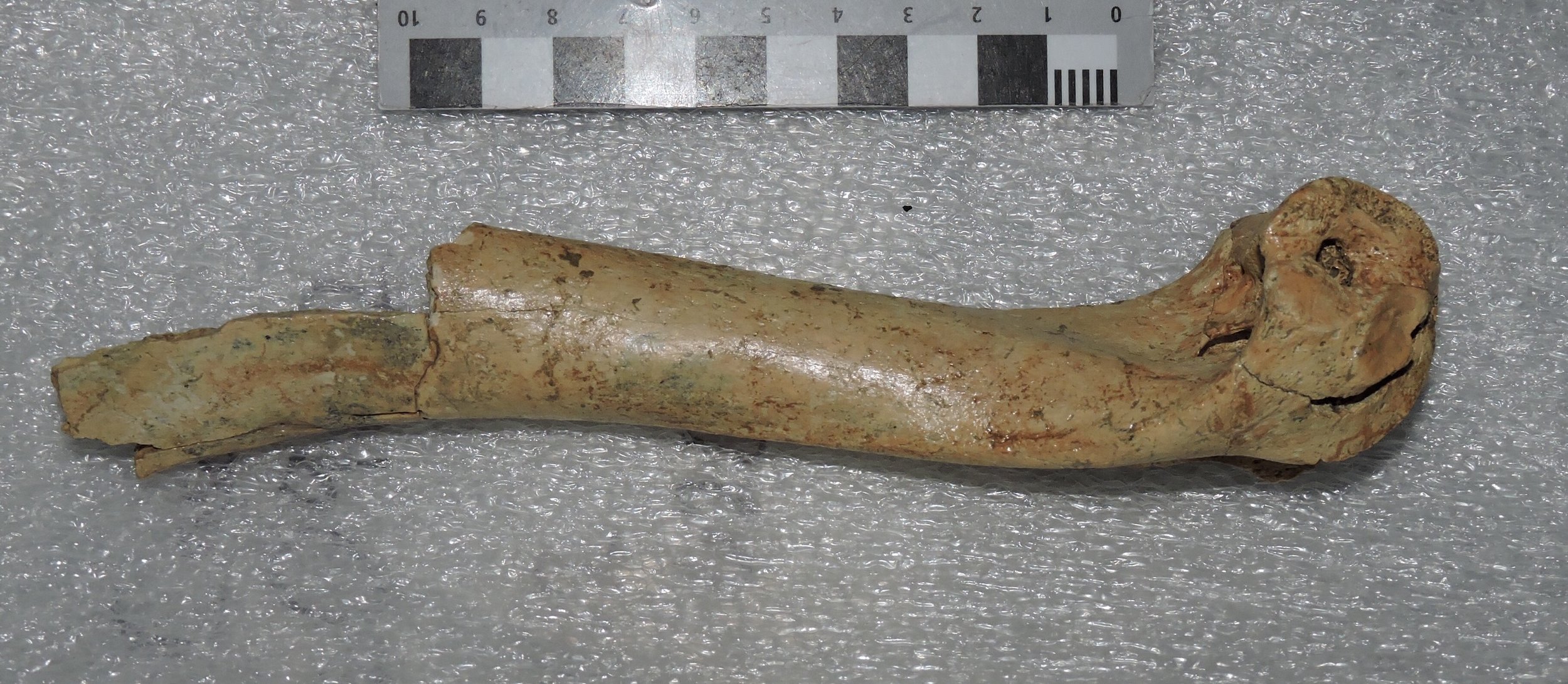 Here's the posterior view:
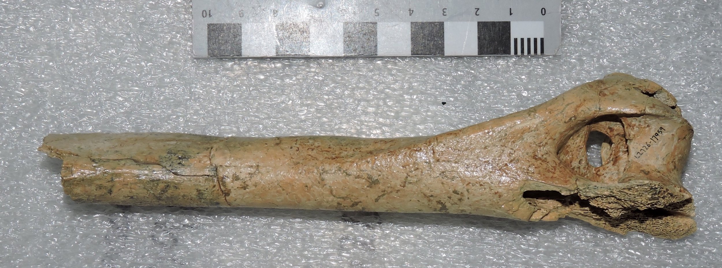 In this view the supratrochlear foramen is sitting in the bottom of a deep semi-circular depression, called the olecranon fossa. When the front leg is held straight, the olecranon process of the ulna (the "point" of the elbow) fits into this depression. This stabilizes the elbow and prevents the upper and lower arms from rotating in opposite directions along their long axes. To see how this works, hold your arm out straight, then rotate your arm like you're turning a screwdriver. Watch your elbow as you do this; you'll see that there's no rotation of the elbow at all, and that all the rotation is occurring either in the forearm and wrist, or at the shoulder.So, moving on from the anatomy, what kind of dog is this? This is actually a pretty large humerus by dog standards, and probably it's too large to be from a fox or a coyote (even the big Pleistocene coyotes). The distal end is damaged on its lateral side, but it looks like the bone was somewhere around 50-55 mm wide at the widest part of the distal end. At that size, this humerus is about right for either a small dire wolf (Canis dirus) or a large grey wolf (Canis lupus). There is other Diamond Valley Lake material that is pretty clearly from Canis dirus, but none that's definitively from Canis lupus (in fact, grey wolf fossils are extremely rare anywhere in California, if they're present at all), so a dire wolf is the more likely identification.
Fossil Friday - colubrids snake vertebra
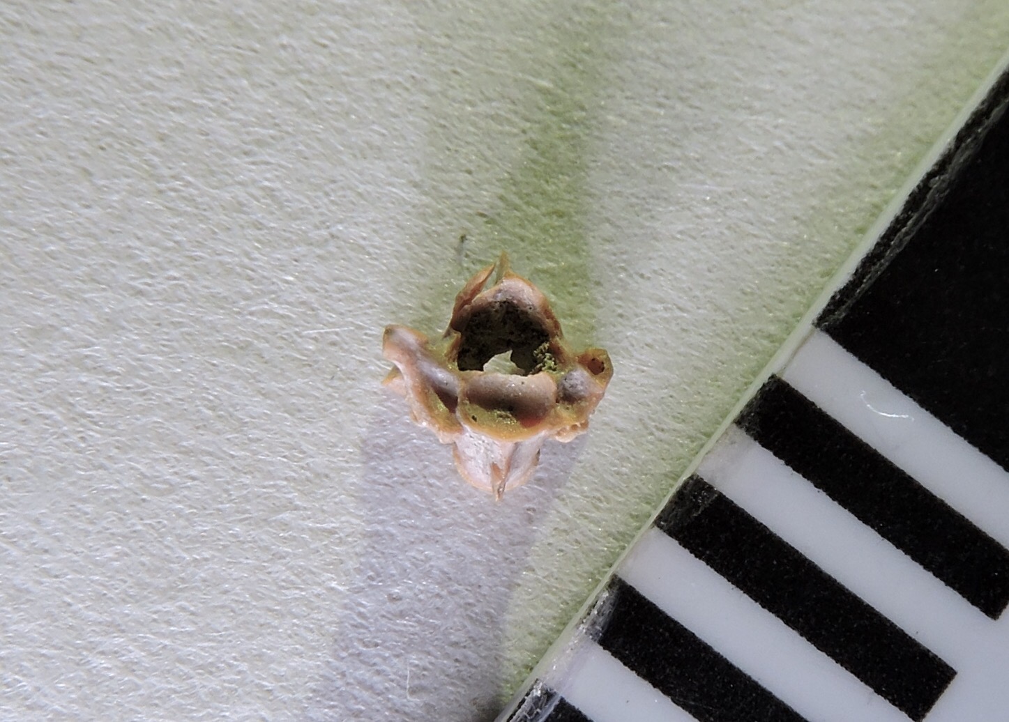 In recognition of World Snake Day (which was yesterday), for today's Fossil Friday we have a Pleistocene snake from Diamond Valley Lake.Reptiles are common in Southern California today, and the same was true during the Pleistocene, including turtles, lizards, and snakes. While rattlesnakes may get more attention, the most diverse and widespread group of snakes are the family Colubridae; in fact, more than half of the extant snake species are colubrids. With such a common group, it's not surprising that their vertebrae turn up fairly commonly as fossils.At the top of the page is a dorsal vertebra, from the region that makes up the bulk of a snake's body. The vertebra itself is about 3 millimeters across at the widest point, but in the living snake a rib would have been attached to each side. It's shown here in close to anterior view, but slightly below and to the right. Like other snakes this vertebra is procoelous, meaning that it articulates with other vertebrae in part through ball-and-socket joints. The socket on the front of the vertebra is visible here; there's a corresponding ball joint on the other end of the vertebra.Colubrids are still diverse in Southern California, and include such diverse forms as king snakes, garter snakes, and gopher snakes (such as Pituophis catanifer, below, photographed on the museum campus). With such a range of closely-related species to choose from, so far we can only say that this vertebra represents some type of colubrid.
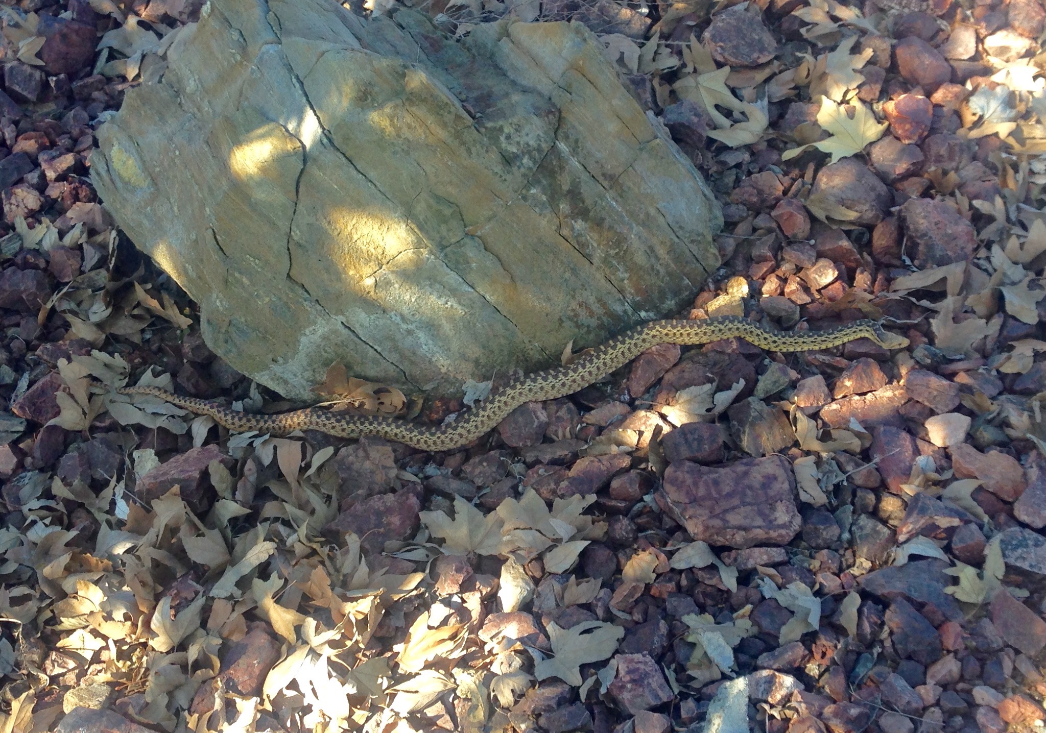
Fossil Friday - quail hand
 When studying comparative anatomy, one of the most basic skills is identifying homologous structures - the same bone or organ in different organisms. This can be tricky, as a structure may become hugely modified in the course of evolution as it is co-opted for new purposes. The bird's wing provides a classic example.Birds' wings are modified forelimbs, and as such have the same basic components as any other tetrapod's forelimb, including a wrist and hand. A typical tetrapod hand commonly has five bones called metacarpals, that articulate at the proximal ends with a series of small bones called the carpals (wrist bones). In birds there are only three metacarpals, a trait they inherited from their theropod dinosaur ancestors. Moreover, most of the bird carpals and the metacarpals are fused together into a single structure called the carpometacarpus.At the top of the page is a partial carpometacarpus from the Pleistocene deposits at Diamond Valley Lake. This represents part of the proximal end (the wrist end) of a small right carpometacarpus (the small scale increments are millimeters). The long shaft is the 2nd metacarpal, the hand bone associated with the index finger. Even in such a small fragment several features are visible, such as those indicated below:
 The projection marked "1" is the 1st metacarpal, the one associated with the thumb; in birds it it reduced and solidly fused to the carpals. The thumb bone, which is called the alula in birds and supports feathers on the leading edge of the wing, articulates at this point.Number "2" is the broken base of the 3rd metacarpal, the one associated with the middle finger. Birds don't have the 4th and 5th metacarpals, the ones associated with the ring and pinky fingers; these were lost in bird ancestors millions of years before the first birds appear in the fossil record.Number "3" is called the intermetacarpal tuberosity; it's a bony spur of metacarpal 2 that projects into the gap between metacarpals 2 and 3. In this specimen, the tuberosity is very large relative to the size of the metacarpal. That's significant, because such a large tuberosity is generally only present in a group of birds called the Galliformes (turkeys, chickens, quails, and their relatives). The size and shape of this particular bone suggests that belongs to a New World quail from the genus Callipepla.Callipepla is still a common bird in southern California, with two different species considered to be native to the area. Callipepla californica is California's official state bird, and C. gambelii is also common in this area (below is C. gambelii from the North Carolina Zoo) . Two other species of Callipepla are found in the southwestern United States or northern Mexico but not currently in California. With such a fragmentary specimen, any of these closely related species are a possibilities for this Diamond Valley Lake specimen. We have a number of Callipepla bones in the collection, suggesting that, like today, quail were a common component of the Diamond Valley Lake fauna during the Pleistocene.